Explore
 Validate
 Learn
LearnMA1-33972
antibody from Invitrogen Antibodies
Targeting: S100A9
60B8AG, CAGB, CFAG, CGLB, LIAG, MAC387, MIF, MRP14, NIF, P14
 Western blot
 Immunohistochemistry
Antibody data
- Antibody Data
- Antigen structure
- References [0]
- Comments [0]
- Validations
- Western blot [1]
- Immunocytochemistry [1]
Submit
Validation data
Reference
Comment
Report error
- Product number
- MA1-33972 - Provider product page

- Provider
- Invitrogen Antibodies
- Product name
- Calprotectin Monoclonal Antibody (MAC387)
- Antibody type
- Monoclonal
- Antigen
- Other
- Description
- For staining of paraffin sections, protein digestion pre-treatment (e.g. trypsin), 0.1% for 10 minutes or antigen retrieval using heat treatment is required. Store product as a concentrated solution. Centrifuge briefly prior to opening the vial.
- Reactivity
- Human, Mouse, Rat, Bovine, Canine, Feline, Goat, Guinea Pig, Rabbit
- Host
- Mouse
- Isotype
- IgG
- Antibody clone number
- MAC387
- Vial size
- 100 µg
- Concentration
- 1 mg/mL
- Storage
- Store at 4°C short term. For long term storage, store at -20°C, avoiding freeze/thaw cycles.
No comments: Submit comment
Supportive validation
- Submitted by
- Invitrogen Antibodies (provider)
- Main image
- Experimental details
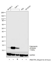
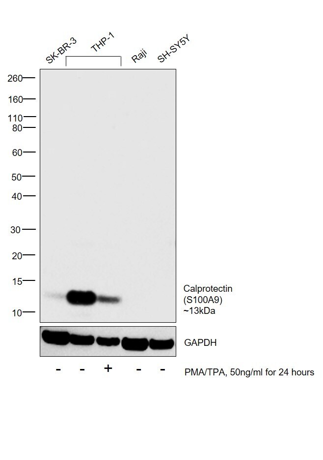
- Western blot was performed using Anti-Calprotectin Monoclonal Antibody (MAC387) (Product # MA1-33972) and a 13kDa band corresponding to Calprotectin was observed in SK-BR-3 and THP-1 but was absent in Raji and SH-SY5Y which are reported to be negative. Expression of Calprotectin was downregulated upon treatment of THP-1 with PMA/TPA [10.1073/pnas.0709958105]. Membrane enriched cell extracts (30 µg lysate) of SK-BR-3 (Lane 1), THP-1 (Lane 2), THP-1 treated with PMA/TPA (50ng/ml for 24 hours) (Lane 3), Raji (Lane 4) and SH-SY5Y (Lane 5) were electrophoresed using Novex® NuPAGE® 4-12 % Bis-Tris gel (Product # NP0322BOX). Resolved proteins were then transferred onto a nitrocellulose membrane (Product # IB23001) by iBlot® 2 Dry Blotting System (Product # IB21001). The blot was probed with the primary antibody (1:1000 dilution) and detected by chemiluminescence with Goat anti-Mouse IgG (H+L), Superclonal™ Recombinant Secondary Antibody, HRP (Product # A28177, 1:4000 dilution) using the iBright FL 1000 (Product # A32752). Chemiluminescent detection was performed using Novex® ECL Chemiluminescent Substrate Reagent Kit (Product # WP20005).
Supportive validation
- Submitted by
- Invitrogen Antibodies (provider)
- Main image
- Experimental details
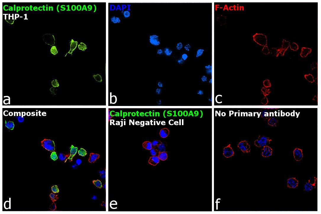
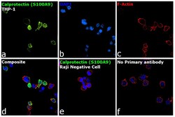
- Immunofluorescence analysis of Calprotectin was performed using log phase THP-1 and Raji cells. The cells were fixed with 4% paraformaldehyde for 10 minutes, permeabilized with 0.1% Triton™ X-100 for 15 minutes, and blocked with 2% BSA for 1 hour at room temperature. The cells were labeled with Calprotectin Monoclonal Antibody (MAC387) (Product # MA1-33972) at 1:250 dilution in 0.1% BSA, incubated at 4 degree Celsius overnight and then labeled with Donkey anti-Mouse IgG (H+L) Highly Cross-Adsorbed Secondary Antibody, Alexa Fluor Plus 488 (Product # A32766) at a dilution of 1:2000 for 45 minutes at room temperature (Panel a: green). Nuclei (Panel b: blue) were stained with SlowFade® Gold Antifade Mountant with DAPI (Product # S36938). F-actin (Panel c: red) was stained with Rhodamine Phalloidin (Product # R415, 1:300). Panel d represents the merged image showing localization to plasma membrane, nucleus and cytoplasm. Panel e shows Raji cells with no expression of Calprotectin. Panel f represents control cells with no primary antibody to assess background. The images were captured at 60X magnification.