Explore
 Validate
 Learn
LearnPA1-46489
antibody from Invitrogen Antibodies
Targeting: S100A9
60B8AG, CAGB, CFAG, CGLB, LIAG, MAC387, MIF, MRP14, NIF, P14
 Western blot
 Immunocytochemistry
Antibody data
- Antibody Data
- Antigen structure
- References [5]
- Comments [0]
- Validations
- Immunocytochemistry [2]
- Other assay [6]
Submit
Validation data
Reference
Comment
Report error
- Product number
- PA1-46489 - Provider product page

- Provider
- Invitrogen Antibodies
- Product name
- S100A9 Polyclonal Antibody
- Antibody type
- Polyclonal
- Antigen
- Other
- Description
- Suggested positive control: antigen standard for S100A9 (transient overexpression lysate), HL-60 + DMSO.
- Reactivity
- Human, Mouse, Rat
- Host
- Rabbit
- Isotype
- IgG
- Vial size
- 100 μL
- Concentration
- Conc. Not Determined
- Storage
- Store at 4°C short term. For long term storage, store at -20°C, avoiding freeze/thaw cycles.
Submitted references Rapamycin-filgrastim combination therapy ameliorates portal hypertension-induced splenomegaly: Role of β actin and S100A9 proteins modulation.
S100A8 and S100A9, both transcriptionally regulated by PU.1, promote epithelial-mesenchymal transformation (EMT) and invasive growth of dermal keratinocytes during scar formation post burn.
Quantitative proteomic characterization of microvesicles/exosomes from the cerebrospinal fluid of patients with acute bilirubin encephalopathy.
Distinct molecular signatures of mild extrinsic and intrinsic atopic dermatitis.
Analysis of protein expression in periodontal pocket tissue: a preliminary study.
Abdelrahman SA, Abdelfatah MM, Keshta AT
Iranian journal of basic medical sciences 2022 Jun;25(6):732-744
Iranian journal of basic medical sciences 2022 Jun;25(6):732-744
S100A8 and S100A9, both transcriptionally regulated by PU.1, promote epithelial-mesenchymal transformation (EMT) and invasive growth of dermal keratinocytes during scar formation post burn.
Xu Z, Cheng C, Kong R, Liu Y, Wang S, Ma Y, Xing X
Aging 2021 Jun 7;13(11):15523-15537
Aging 2021 Jun 7;13(11):15523-15537
Quantitative proteomic characterization of microvesicles/exosomes from the cerebrospinal fluid of patients with acute bilirubin encephalopathy.
Tan N, Hu S, Hu Z, Wu Z, Wang B
Molecular medicine reports 2020 Aug;22(2):1257-1268
Molecular medicine reports 2020 Aug;22(2):1257-1268
Distinct molecular signatures of mild extrinsic and intrinsic atopic dermatitis.
Martel BC, Litman T, Hald A, Norsgaard H, Lovato P, Dyring-Andersen B, Skov L, Thestrup-Pedersen K, Skov S, Skak K, Poulsen LK
Experimental dermatology 2016 Jun;25(6):453-9
Experimental dermatology 2016 Jun;25(6):453-9
Analysis of protein expression in periodontal pocket tissue: a preliminary study.
Monari E, Cuoghi A, Bellei E, Bergamini S, Lucchi A, Tomasi A, Cortellini P, Zaffe D, Bertoldi C
Proteome science 2015;13:33
Proteome science 2015;13:33
No comments: Submit comment
Supportive validation
- Submitted by
- Invitrogen Antibodies (provider)
- Main image

- Experimental details
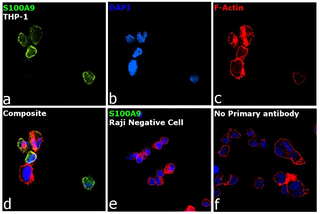
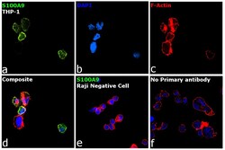
- Immunofluorescence analysis of S100A9 was performed using log phase THP-1 and Raji cells. The cells were fixed with 4% paraformaldehyde for 10 minutes, permeabilized with 0.1% Triton™ X-100 for 15 minutes, and blocked with 2% BSA for 1 hour at room temperature. The cells were labeled with S100A9 Polyclonal Antibody (Product # PA1-46489) at 1:200 dilution in 0.1% BSA, incubated at 4 degree Celsius overnight and then labeled with Donkey anti-Rabbit IgG (H+L) Highly Cross-Adsorbed Secondary Antibody, Alexa Fluor Plus 488 (Product # A32790) at a dilution of 1:2000 for 45 minutes at room temperature (Panel a: green). Nuclei (Panel b: blue) were stained with SlowFade® Gold Antifade Mountant with DAPI (Product # S36938). F-actin (Panel c: red) was stained with Rhodamine Phalloidin (Product # R415, 1:300). Panel d represents the merged image showing localization to plasma membrane, nucleus and cytoplasm. Panel e shows Raji cells with no expression of S100A9. Panel f represents control cells with no primary antibody to assess background. The images were captured at 60X magnification.
- Submitted by
- Invitrogen Antibodies (provider)
- Main image
- Experimental details
- Immunofluorescence analysis of S100A9 was performed using log phase THP-1 and Raji cells. The cells were fixed with 4% paraformaldehyde for 10 minutes, permeabilized with 0.1% Triton™ X-100 for 15 minutes, and blocked with 2% BSA for 1 hour at room temperature. The cells were labeled with S100A9 Polyclonal Antibody (Product # PA1-46489) at 1:200 dilution in 0.1% BSA, incubated at 4 degree Celsius overnight and then labeled with Donkey anti-Rabbit IgG (H+L) Highly Cross-Adsorbed Secondary Antibody, Alexa Fluor Plus 488 (Product # A32790) at a dilution of 1:2000 for 45 minutes at room temperature (Panel a: green). Nuclei (Panel b: blue) were stained with SlowFade® Gold Antifade Mountant with DAPI (Product # S36938). F-actin (Panel c: red) was stained with Rhodamine Phalloidin (Product # R415, 1:300). Panel d represents the merged image showing localization to plasma membrane, nucleus and cytoplasm. Panel e shows Raji cells with no expression of S100A9. Panel f represents control cells with no primary antibody to assess background. The images were captured at 60X magnification.

Supportive validation
- Submitted by
- Invitrogen Antibodies (provider)
- Main image
- Experimental details
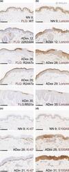
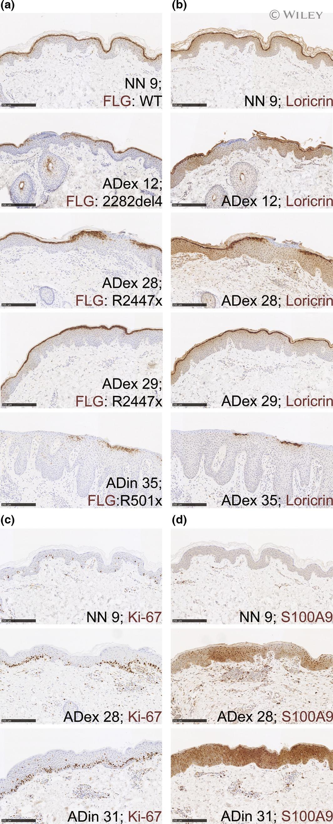
- Histological analysis of proteins involved in epidermal barrier function. Immunohistochemical staining (brown) of filaggrin, loricrin, Ki-67 and S100A9 in skin biopsies from patients with extrinsic atopic dermatitis ( AD ex), intrinsic AD ( AD in) and healthy controls ( NN ). (a) Filaggrin staining of skin samples from NN and patients with a detected mutation in FLG at the indicated loci: 2282del4, R2447x or R501x. (b) Loricrin staining of skin samples from NN and patients with a mutation in FLG . (c) Ki-67 staining of skin samples from NN and patients with extrinsic and intrinsic AD , respectively. (d) S100A9 staining of skin samples from NN and patients with extrinsic and intrinsic AD , respectively. Numbers correspond to the ID of the specific subjects as in Table S1. Scale bars = 250 mu m.
- Submitted by
- Invitrogen Antibodies (provider)
- Main image

- Experimental details
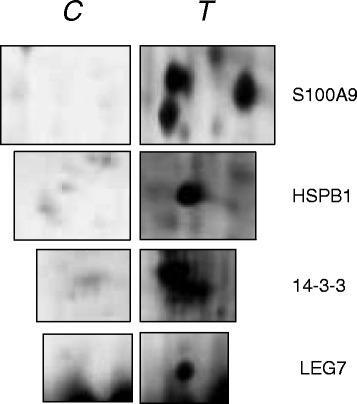
- Fig. 3 Detailed protein spots, differentially expressed in C and T specimens
- Submitted by
- Invitrogen Antibodies (provider)
- Main image
- Experimental details
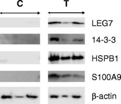
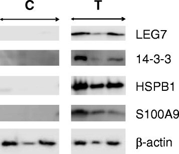
- Fig. 4 Representative images of Western blot analyses in C and T specimens for LEG7, 14-3-3, HSPB1, S100A9 and beta-actin. The beta-actin was utilized as reference for protein samples loading and integrity
- Submitted by
- Invitrogen Antibodies (provider)
- Main image
- Experimental details
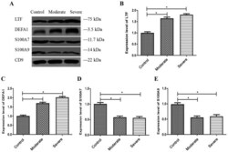
- Figure 6. Verification of differentially expressed proteins using western blotting. (A) Representative western blots demonstrate the expression levels of LTF, DEFA1, S100A7 and S100A9 in the MV/E isolated from the CSF. Histogram data shows the densitometric semi-quantification (mean +- SD) of (B) LTF, (C) DEFA1, (D) S100A7 and (E) S100A9 expression levels. Protein densitometric semi-quantification was normalized to the density of CD9 protein. CD9 is an exosome surface marker. *P

- Submitted by
- Invitrogen Antibodies (provider)
- Main image
- Experimental details
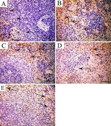
- Figure 10 S100A9 immunostained section in the spleen showing A: Control group shows positive cytoplasmic immunoreaction for S100 antibody in some cells of the red pulp (arrow). B: TAA group showing increased cytoplasmic immunoreaction in cells of red and white pulp (arrow). C: Rapamycin-treated group showing increased cytoplasmic immunoreaction in cells of red pulp (arrow). D: Filgrastim-treated group showing positive cytoplasmic immunoreaction in some cells of the red pulp (arrow). E: Combined rapamycin and filgrastim-treated group showing positive reaction in few cells of red pulp (arrow) (Immunoperoxidase technique for S100 x 40, scale bar 30 um) TTA: thioacetamide

- Submitted by
- Invitrogen Antibodies (provider)
- Main image
- Experimental details
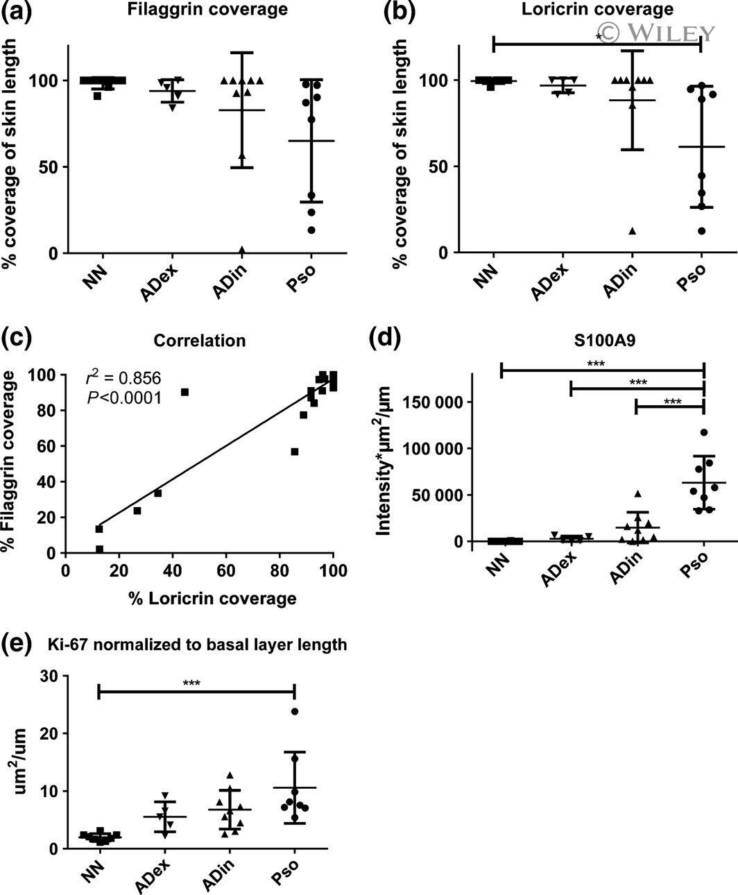
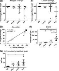
- Different expression of proteins involved in epidermal barrier function. (a, b) Filaggrin and loricrin coverage, respectively. Coverage was determined manually by subtracting the length of the unstained part of the epidermis from the entire length of the epidermis. (c) Correlation of filaggrin and loricrin coverage. (d) Intensity of S100A9 staining multiplied by area and normalized to the length of the skin biopsy. (e) Area of Ki-67 staining normalized to the length of the basal layer. n ( AD ex) = 5; n ( AD in) = 8; n (Pso) = 8; n ( NN ) = 8. Horizontal bars: mean with SD . * P < 0.05; *** P < 0.001. Comparison of groups: 1-way ANOVA with Tukey's multiple comparison test.