Explore
 Validate
 Learn
Learn Western blot
Antibody data
- Antibody Data
- Antigen structure
- References [4]
- Comments [0]
- Validations
- Western blot [2]
- Immunocytochemistry [1]
- Immunohistochemistry [1]
Submit
Validation data
Reference
Comment
Report error
- Product number
- GTX113389 - Provider product page

- Provider
- GeneTex
- Proper citation
- GeneTex Cat#GTX113389, RRID:AB_2036283
- Product name
- Aspartoacylase antibody [N1C3-2]
- Antibody type
- Polyclonal
- Reactivity
- Human, Mouse, Simian
- Host
- Rabbit
Submitted references Spatiotemporal distribution of fibrinogen in marmoset and human inflammatory demyelination.
Gata6 regulates aspartoacylase expression in resident peritoneal macrophages and controls their survival.
N-acetylaspartate (NAA) and N-acetylaspartylglutamate (NAAG) promote growth and inhibit differentiation of glioma stem-like cells.
Expression of aspartoacylase (ASPA) and Canavan disease.
Lee NJ, Ha SK, Sati P, Absinta M, Luciano NJ, Lefeuvre JA, Schindler MK, Leibovitch EC, Ryu JK, Petersen MA, Silva AC, Jacobson S, Akassoglou K, Reich DS
Brain : a journal of neurology 2018 Jun 1;141(6):1637-1649
Brain : a journal of neurology 2018 Jun 1;141(6):1637-1649
Gata6 regulates aspartoacylase expression in resident peritoneal macrophages and controls their survival.
Gautier EL, Ivanov S, Williams JW, Huang SC, Marcelin G, Fairfax K, Wang PL, Francis JS, Leone P, Wilson DB, Artyomov MN, Pearce EJ, Randolph GJ
The Journal of experimental medicine 2014 Jul 28;211(8):1525-31
The Journal of experimental medicine 2014 Jul 28;211(8):1525-31
N-acetylaspartate (NAA) and N-acetylaspartylglutamate (NAAG) promote growth and inhibit differentiation of glioma stem-like cells.
Long PM, Moffett JR, Namboodiri AM, Viapiano MS, Lawler SE, Jaworski DM
The Journal of biological chemistry 2013 Sep 6;288(36):26188-200
The Journal of biological chemistry 2013 Sep 6;288(36):26188-200
Expression of aspartoacylase (ASPA) and Canavan disease.
Sommer A, Sass JO
Gene 2012 Sep 1;505(2):206-10
Gene 2012 Sep 1;505(2):206-10
No comments: Submit comment
Supportive validation
- Submitted by
- GeneTex (provider)
- Main image

- Experimental details
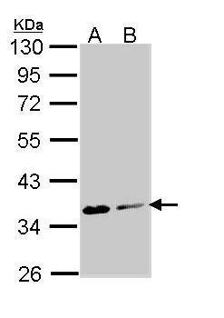
- Sample (30 ?g of whole cell lysate) A: Molt-4 (GTX27912) B: Raji 10% SDS PAGE GTX113389 diluted at 1:1000 The HRP-conjugated anti-rabbit IgG antibody (GTX213110-01) was used to detect the primary antibody.
- Submitted by
- GeneTex (provider)
- Main image
- Experimental details
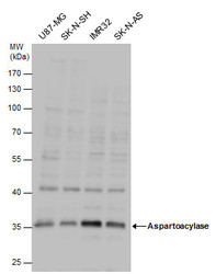
- Aspartoacylase antibody detects Aspartoacylase protein by western blot analysis. Various whole cell extracts (30 ?g) were separated by 10% SDS-PAGE, and the membrane was blotted with Aspartoacylase antibody (GTX113389) diluted by 1:1000. The HRP-conjugated anti-rabbit IgG antibody (GTX213110-01) was used to detect the primary antibody.

Supportive validation
- Submitted by
- GeneTex (provider)
- Main image
- Experimental details
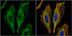
- Aspartoacylase antibody [N1C3-2] detects Aspartoacylase protein at cytoplasm by immunofluorescent analysis.Sample: HeLa cells were fixed in 4% paraformaldehyde at RT for 15 min.Green: Aspartoacylase protein stained by Aspartoacylase antibody [N1C3-2] (GTX113389) diluted at 1:500.Red: alpha Tubulin, a cytoskeleton marker, stained by alpha Tubulin antibody [GT114] (GTX628802) diluted at 1:1000.Blue: Hoechst 33342 staining.

Supportive validation
- Submitted by
- GeneTex (provider)
- Main image
- Experimental details
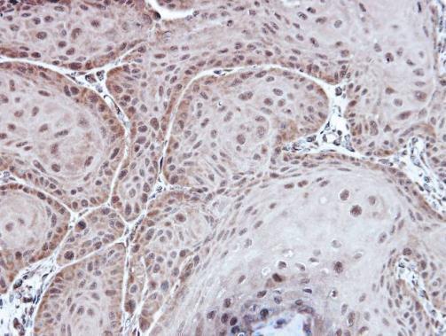
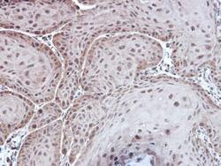
- Immunohistochemical analysis of paraffin-embedded Cal27 xenograft, using Aspartoacylase(GTX113389) antibody at 1:100 dilution.