Explore
 Validate
 Learn
Learn Western blot
 Flow cytometry
Antibody data
- Antibody Data
- Antigen structure
- References [0]
- Comments [0]
- Validations
- Western blot [2]
- Immunocytochemistry [1]
- Immunohistochemistry [9]
- Other assay [1]
Submit
Validation data
Reference
Comment
Report error
- Product number
- UM500006 - Provider product page

- Provider
- OriGene
- Proper citation
- OriGene Cat#UM500006, RRID:AB_2629021
- Product name
- Anti-CD2 mouse monoclonal antibody,clone UMAB6
- Antibody type
- Monoclonal
- Description
- Anti-CD2 mouse monoclonal antibody,clone UMAB6
- Host
- Mouse
- Conjugate
- Unconjugated
- Epitope
- CD2
- Isotype
- IgG
- Antibody clone number
- UMAB6
- Vial size
- 100 µl
- Concentration
- 1.00mg/ml
No comments: Submit comment
Supportive validation
- Submitted by
- OriGene (provider)
- Main image
- Experimental details
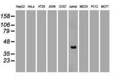
- Western blot analysis of extracts (35ug) from 9 different cell lines by using anti-CD2 monoclonal antibody (Clone UMAB6).
- Validation comment
- WB

- Submitted by
- OriGene (provider)
- Main image
- Experimental details
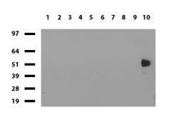
- Western blot of human tissue lysates (15ug) from 10 different tissues (1: Testis, 2: Omentum, 3: Uterus, 4: Breast, 5: Brain, 6: Liver, 7: Ovary, 8: Thyroid 9: Colon, 10: Spleen). Diluation: 1:500.
- Validation comment
- WB
Supportive validation
- Submitted by
- OriGene (provider)
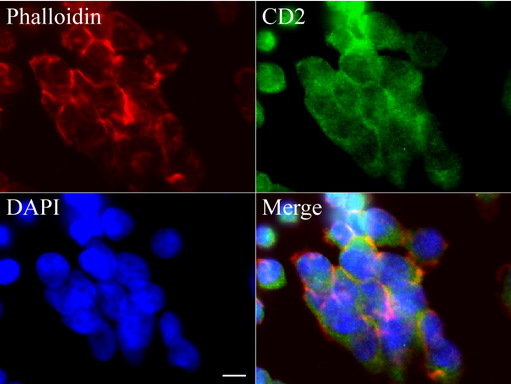
- Main image
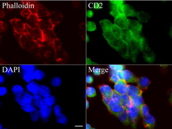
- Experimental details
- Immunofluorescent staining of Jurkat cells using anti-CD2 mouse monoclonal antibody (UM500006, green, 1:100). Actin filaments were labeled with Alexa Fluor? 594 Phalloidin (red), and nuclear with DAPI (blue). Scale bar, 8?m.
- Validation comment
- IF
Supportive validation
- Submitted by
- OriGene (provider)
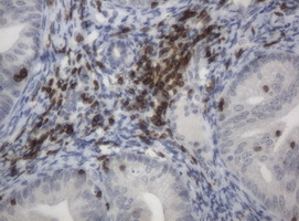
- Main image

- Experimental details
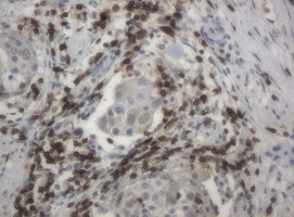
- Immunohistochemical staining of paraffin-embedded Adenocarcinoma of Human endometrium tissue using anti-CD2 mouse monoclonal antibody. (Clone UMAB6, dilution 1:100; heat-induced epitope retrieval by 10mM citric buffer, pH6.0, 120C for 3min)
- Validation comment
- IHC
- Submitted by
- OriGene (provider)
- Main image
- Experimental details
- Immunohistochemical staining of paraffin-embedded Carcinoma of Human bladder tissue using anti-CD2 mouse monoclonal antibody. (Clone UMAB6, dilution 1:100; heat-induced epitope retrieval by 10mM citric buffer, pH6.0, 120C for 3min)
- Validation comment
- IHC
- Submitted by
- OriGene (provider)
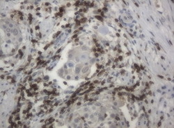
- Main image
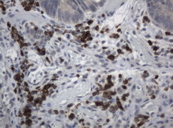
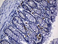
- Experimental details
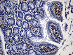
- Immunohistochemical staining of paraffin-embedded Adenocarcinoma of colon tissue using anti-CD2 mouse monoclonal antibody. (Clone UMAB6, dilution 1:100; heat-induced epitope retrieval by 10mM citric buffer, pH6.0, 120C for 3min)
- Validation comment
- IHC
- Submitted by
- OriGene (provider)
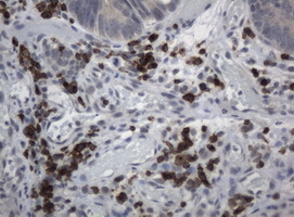
- Main image
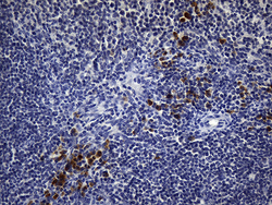
- Experimental details
- Immunohistochemical staining of paraffin-embedded Human lymphoma tissue using anti-CD2 mouse monoclonal antibody. (Clone UMAB6, dilution 1:100; heat-induced epitope retrieval by 10mM citric buffer, pH6.0, 120C for 3min)
- Validation comment
- IHC
- Submitted by
- OriGene (provider)
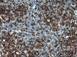
- Main image

- Experimental details
- IHC staining of paraffin-embedded human tonsil using anti-CD2 clone UMAB6 mouse monoclonal antibody at 1:200 of 0.6mg/mL and detection with Polink2 Broad HRP DAB. UM500006 requires heat-induced epitope retrieval with Accel pH8.7 at 95-100C 30 minutes [do not let boil] or 10 min in pressure cooker. The image shows strong membranous and cytoplasmic staining in >50 % of non germinal center cells of tonsil and
- Validation comment
- IHC
- Submitted by
- OriGene (provider)
- Main image
- Experimental details
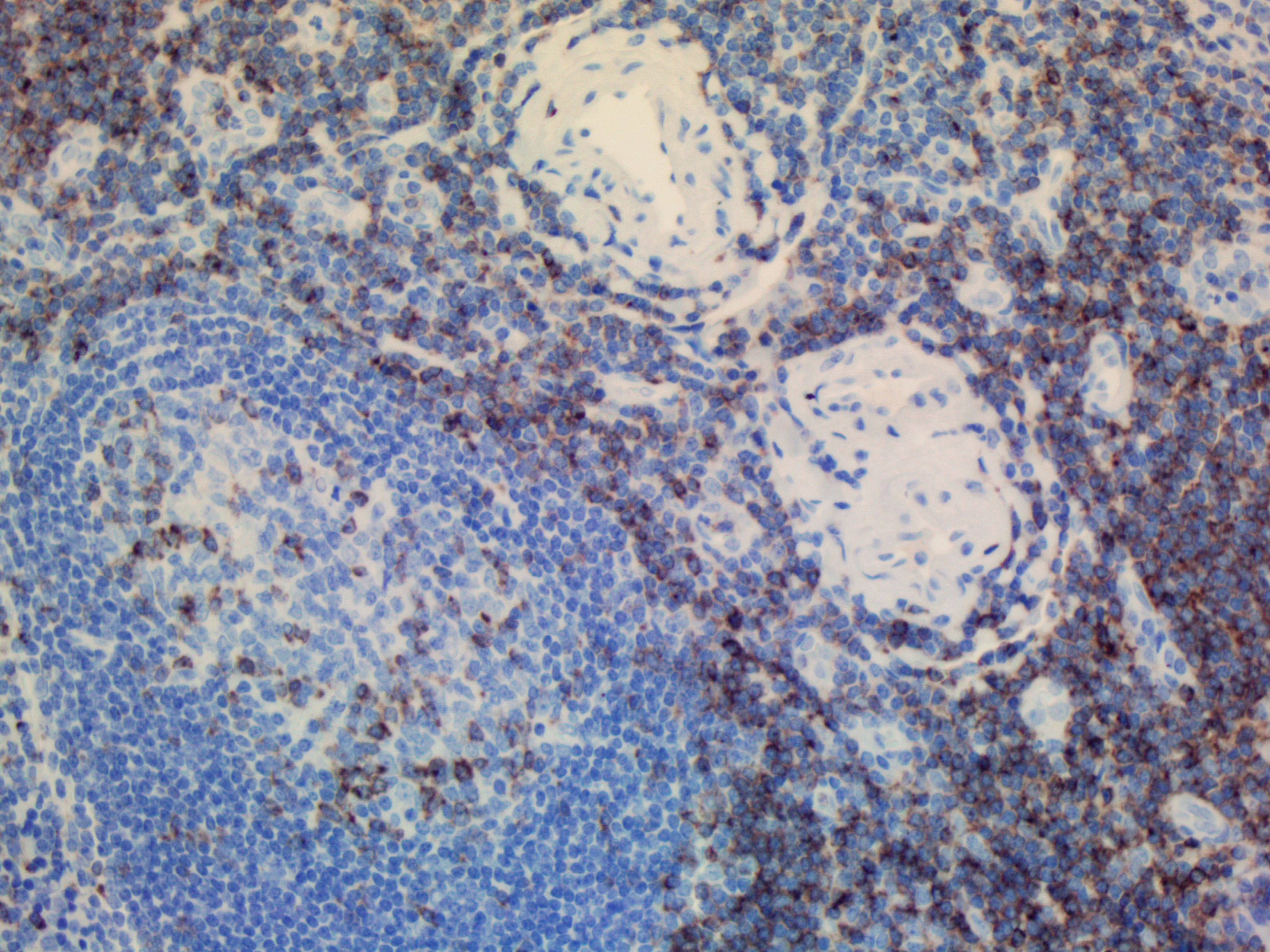
- Immunohistochemical staining of paraffin-embedded mouse spleen tissue using anti-CD2 clone UMAB6 mouse monoclonal antibody. (Heat-induced epitope retrieval by 1mM EDTA in 10mM Tris buffer (pH8.5) at 120 oC for 3 min, UM500006)(1:500).
- Validation comment
- IHC

- Submitted by
- OriGene (provider)
- Main image
- Experimental details
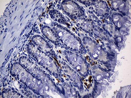
- Immunohistochemical staining of paraffin-embedded mouse colon tissue using anti-CD2 clone UMAB6 mouse monoclonal antibody. (Heat-induced epitope retrieval by 1mM EDTA in 10mM Tris buffer (pH8.5) at 120°C for 3 min, UM500006)(1:500).
- Validation comment
- IHC
- Submitted by
- OriGene (provider)
- Main image
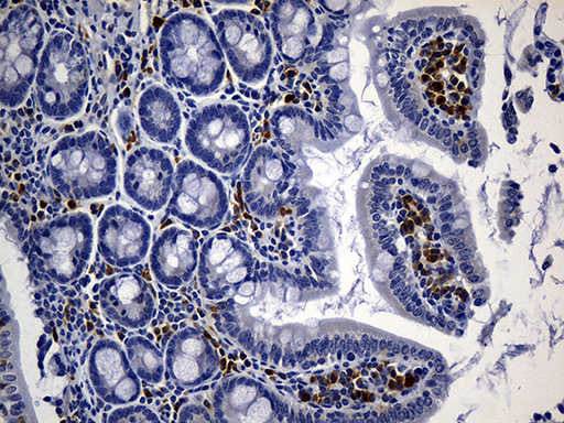
- Experimental details
- Immunohistochemical staining of paraffin-embedded mouse ascending colon tissue anti-CD2 clone UMAB6 mouse monoclonal antibody. (Heat-induced epitope retrieval by 1mM EDTA in 10mM Tris buffer (pH8.5) at 120°C for 3 min, UM500006)(1:500).
- Validation comment
- IHC
- Submitted by
- OriGene (provider)
- Main image
- Experimental details
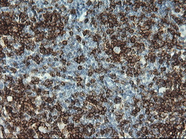
- IHC staining of FFPE human spleen using anti-CD2 clone UMAB6 mouse monoclonal antibody at 1:200 and detection with Polink2 Broad HRP DAB. UM500006 requires heat-induced epitope retrieval with Citrate pH6.O. The image shows strong membranous and cytoplasmic staining.
- Validation comment
- IHC

Supportive validation
- Submitted by
- OriGene (provider)
- Main image
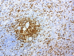
- Experimental details
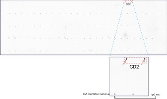
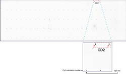
- OriGene overexpression protein microarray chip was immunostained with UltraMAB anti-CD2 mouse monoclonal antibody (Clone UMAB6). The positive reactive proteins are highlighted with two red arrows in the enlarged subarray. All the positive controls spotted in this subarray are also labeled for clarification. These data show that UltraMAB anti-CD2 (Clone UMAB6) very specifically recognizes Cd2 antigen on OriGene protein microarray chip.
- Validation comment
- 10K-CHIP