Explore
 Validate
 Learn
Learn Flow cytometry
Antibody data
- Antibody Data
- Antigen structure
- References [7]
- Comments [0]
- Validations
- Flow cytometry [1]
- Other assay [1]
Submit
Validation data
Reference
Comment
Report error
- Product number
- 12-0029-42 - Provider product page

- Provider
- Invitrogen Antibodies
- Product name
- CD2 Monoclonal Antibody (RPA-2.10), PE, eBioscience™
- Antibody type
- Monoclonal
- Antigen
- Other
- Description
- Description: The RPA-2.10 monoclonal antibody reacts with human CD2, a 50 kDa cell surface receptor expressed by a majority of thymocytes, all mature T cells and subset of NK cells. CD2 is a ligand for CD58 in the human and is involved in adhesion and activation of T cells. RPA-2.10 blocks mixed lymphocyte reaction.
- Conjugate
- Yellow dye
- Antibody clone number
- RPA-2.10
- Concentration
- 5 µL/Test
Submitted references Evaluation of Canonical Inflammasome Activation in Human Monocytes by Imaging Flow Cytometry.
Desynchronization of the molecular clock contributes to the heterogeneity of the inflammatory response.
Constraints on GPCR Heterodimerization Revealed by the Type-4 Induced-Association BRET Assay.
Signaling pathways activated by a protease allergen in basophils.
Forced expression of cyclin-dependent kinase 6 confers resistance of pro-B acute lymphocytic leukemia to Gleevec treatment.
A monoclonal antibody selection for immunohistochemical examination of lymphoid tissues from non-human primates.
Position effect variegation and imprinting of transgenes in lymphocytes.
Lage SL, Dominical VM, Wong CS, Sereti I
Frontiers in immunology 2019;10:1284
Frontiers in immunology 2019;10:1284
Desynchronization of the molecular clock contributes to the heterogeneity of the inflammatory response.
Allen NC, Philip NH, Hui L, Zhou X, Franklin RA, Kong Y, Medzhitov R
Science signaling 2019 Mar 5;12(571)
Science signaling 2019 Mar 5;12(571)
Constraints on GPCR Heterodimerization Revealed by the Type-4 Induced-Association BRET Assay.
Felce JH, MacRae A, Davis SJ
Biophysical journal 2019 Jan 8;116(1):31-41
Biophysical journal 2019 Jan 8;116(1):31-41
Signaling pathways activated by a protease allergen in basophils.
Rosenstein RK, Bezbradica JS, Yu S, Medzhitov R
Proceedings of the National Academy of Sciences of the United States of America 2014 Nov 18;111(46):E4963-71
Proceedings of the National Academy of Sciences of the United States of America 2014 Nov 18;111(46):E4963-71
Forced expression of cyclin-dependent kinase 6 confers resistance of pro-B acute lymphocytic leukemia to Gleevec treatment.
Kuo TC, Chavarria-Smith JE, Huang D, Schlissel MS
Molecular and cellular biology 2011 Jul;31(13):2566-76
Molecular and cellular biology 2011 Jul;31(13):2566-76
A monoclonal antibody selection for immunohistochemical examination of lymphoid tissues from non-human primates.
Kap YS, van Meurs M, van Driel N, Koopman G, Melief MJ, Brok HP, Laman JD, 't Hart BA
The journal of histochemistry and cytochemistry : official journal of the Histochemistry Society 2009 Dec;57(12):1159-67
The journal of histochemistry and cytochemistry : official journal of the Histochemistry Society 2009 Dec;57(12):1159-67
Position effect variegation and imprinting of transgenes in lymphocytes.
Williams A, Harker N, Ktistaki E, Veiga-Fernandes H, Roderick K, Tolaini M, Norton T, Williams K, Kioussis D
Nucleic acids research 2008 Apr;36(7):2320-9
Nucleic acids research 2008 Apr;36(7):2320-9
No comments: Submit comment
Supportive validation
- Submitted by
- Invitrogen Antibodies (provider)
- Main image
- Experimental details
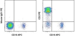
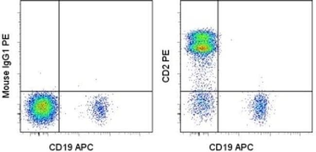
- Staining of normal human peripheral blood cells with Anti-Human CD19 APC (Product # 17-0199-42) and Mouse IgG1 K Isotype Control PE (Product # 12-4714-81) (left) or Anti-Human/Non-Human Primate CD2 PE (right). Cells in the lymphocyte gate were used for analysis.
- Conjugate
- Yellow dye
Supportive validation
- Submitted by
- Invitrogen Antibodies (provider)
- Main image
- Experimental details
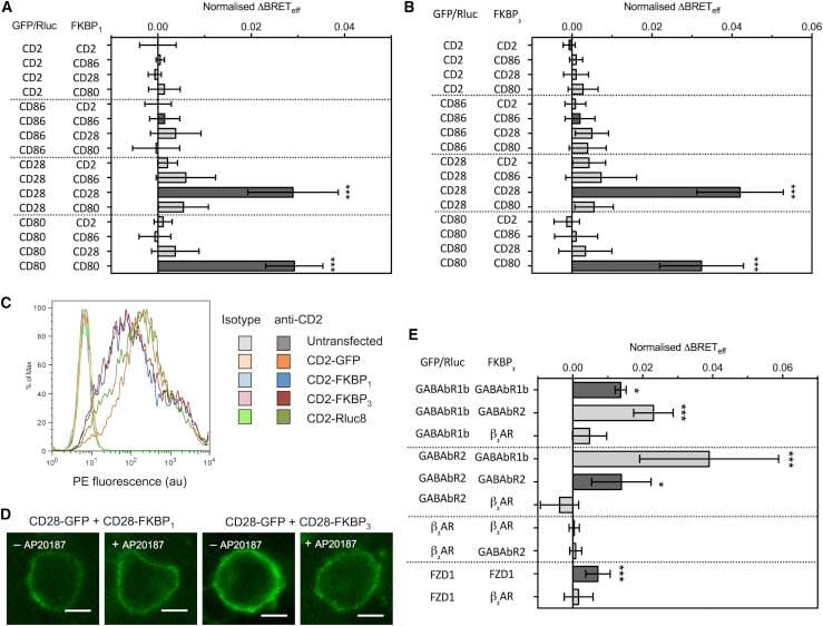
- Figure 2 Type-4 BRET assay applied to known controls. ( A ) The mean Delta BRET eff of type-1 transmembrane protein controls using the FKBP 1 type-4 BRET assay. Only the known dimers CD28 and CD80 exhibited significantly nonzero Delta BRET eff when expressed as homomeric pairs. Dark gray bars indicate like-like pairs. ( B ) The mean Delta BRET eff of type-1 transmembrane protein controls using the FKBP 3 type-4 BRET assay. Delta BRET eff is larger for CD28 and CD80 than with the FKBP 1 approach. ( C ) A histogram of CD2 expression in HEK-293T cells transfected with 1 mu g of differently tagged forms of CD2, as measured by flow cytometry. ( D ) Confocal microscopy of CD28-GFP coexpressed with CD28-FKBP 1/3 in the presence and absence of AP20187. No clear GFP internalization was evident. ( E ) The mean Delta BRET eff of GPCRs of known stoichiometry using the FKBP 3 type-4 BRET assay. For all panels, bars indicate mean +- SD. Probability is indicated for difference from Delta BRET eff = 0; * p < 0.05, *** p < 0.005. All data are the result of n >= 3 independent experiments.
- Conjugate
- Yellow dye