Explore
 Validate
 Learn
Learn Flow cytometry
Antibody data
- Antibody Data
- Antigen structure
- References [4]
- Comments [0]
- Validations
- Flow cytometry [1]
- Other assay [1]
Submit
Validation data
Reference
Comment
Report error
- Product number
- 15-0029-42 - Provider product page

- Provider
- Invitrogen Antibodies
- Product name
- CD2 Monoclonal Antibody (RPA-2.10), PE-Cyanine5, eBioscience™
- Antibody type
- Monoclonal
- Antigen
- Other
- Description
- Description: The RPA-2.10 monoclonal antibody reacts with human CD2, a 50 kDa cell surface receptor expressed by a majority of thymocytes, all mature T cells and subset of NK cells. CD2 is a ligand for CD58 in the human and is involved in adhesion and activation of T cells. RPA-2.10 blocks mixed lymphocyte reaction. RPA-2.10 crossreacts to non-human primates and pigs. Applications Reported: This RPA-2.10 antibody has been reported for use in flow cytometric analysis. Applications Tested: This RPA-2.10 antibody has been pre-titrated and tested by flow cytometric analysis of normal human peripheral blood cells. This can be used at 5 µL (0.06 µg) per test. A test is defined as the amount (µg) of antibody that will stain a cell sample in a final volume of 100 µL. Cell number should be determined empirically but can range from 10^5 to 10^8 cells/test. Light sensitivity: This tandem dye is sensitive photo-induced oxidation. Please protect this vial and stained samples from light. Fixation: Samples can be stored in IC Fixation Buffer (Product # 00-8222) (100 µL cell sample + 100 µL IC Fixation Buffer) or 1-step Fix/Lyse Solution (Product # 00-5333) for up to 3 days in the dark at 4°C with minimal impact on brightness and FRET efficiency/compensation. Some generalizations regarding fluorophore performance after fixation can be made, but clone specific performance should be determined empirically. Excitation: 488-561 nm; Emission: 667 nm; Laser: Blue Laser, Green Laser, Yellow-Green Laser. Filtration: 0.2 µm post-manufacturing filtered.
- Reactivity
- Human, Porcine
- Host
- Mouse
- Isotype
- IgG
- Antibody clone number
- RPA-2.10
- Vial size
- 100 Tests
- Concentration
- 5 µL/Test
- Storage
- 4° C, store in dark, DO NOT FREEZE!
Submitted references Ultra-High-Frequency Reprogramming of Individual Long-Term Hematopoietic Stem Cells Yields Low Somatic Variant Induced Pluripotent Stem Cells.
Constraints on GPCR Heterodimerization Revealed by the Type-4 Induced-Association BRET Assay.
New IDH1 mutant inhibitors for treatment of acute myeloid leukemia.
A monoclonal antibody selection for immunohistochemical examination of lymphoid tissues from non-human primates.
Wang K, Guzman AK, Yan Z, Zhang S, Hu MY, Hamaneh MB, Yu YK, Tolu S, Zhang J, Kanavy HE, Ye K, Bartholdy B, Bouhassira EE
Cell reports 2019 Mar 5;26(10):2580-2592.e7
Cell reports 2019 Mar 5;26(10):2580-2592.e7
Constraints on GPCR Heterodimerization Revealed by the Type-4 Induced-Association BRET Assay.
Felce JH, MacRae A, Davis SJ
Biophysical journal 2019 Jan 8;116(1):31-41
Biophysical journal 2019 Jan 8;116(1):31-41
New IDH1 mutant inhibitors for treatment of acute myeloid leukemia.
Okoye-Okafor UC, Bartholdy B, Cartier J, Gao EN, Pietrak B, Rendina AR, Rominger C, Quinn C, Smallwood A, Wiggall KJ, Reif AJ, Schmidt SJ, Qi H, Zhao H, Joberty G, Faelth-Savitski M, Bantscheff M, Drewes G, Duraiswami C, Brady P, Groy A, Narayanagari SR, Antony-Debre I, Mitchell K, Wang HR, Kao YR, Christopeit M, Carvajal L, Barreyro L, Paietta E, Makishima H, Will B, Concha N, Adams ND, Schwartz B, McCabe MT, Maciejewski J, Verma A, Steidl U
Nature chemical biology 2015 Nov;11(11):878-86
Nature chemical biology 2015 Nov;11(11):878-86
A monoclonal antibody selection for immunohistochemical examination of lymphoid tissues from non-human primates.
Kap YS, van Meurs M, van Driel N, Koopman G, Melief MJ, Brok HP, Laman JD, 't Hart BA
The journal of histochemistry and cytochemistry : official journal of the Histochemistry Society 2009 Dec;57(12):1159-67
The journal of histochemistry and cytochemistry : official journal of the Histochemistry Society 2009 Dec;57(12):1159-67
No comments: Submit comment
Supportive validation
- Submitted by
- Invitrogen Antibodies (provider)
- Main image

- Experimental details
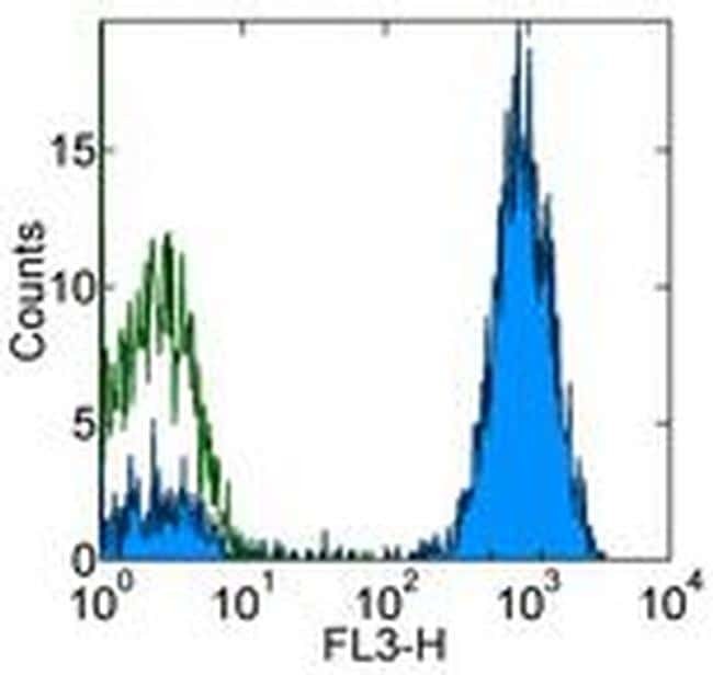
- Staining of normal human peripheral blood cells with Mouse IgG1 K Isotype Control PE-Cyanine5 (Product # 15-4714-81) (open histogram) or Anti-Human CD2 PE-Cyanine5 (filled histogram). Total cells were used for analysis.
Supportive validation
- Submitted by
- Invitrogen Antibodies (provider)
- Main image
- Experimental details
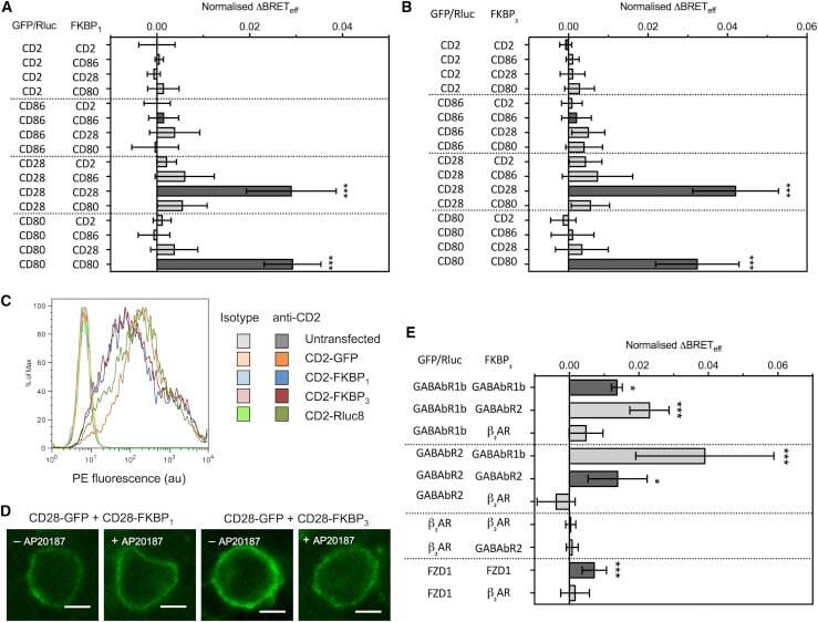
- Figure 2 Type-4 BRET assay applied to known controls. ( A ) The mean Delta BRET eff of type-1 transmembrane protein controls using the FKBP 1 type-4 BRET assay. Only the known dimers CD28 and CD80 exhibited significantly nonzero Delta BRET eff when expressed as homomeric pairs. Dark gray bars indicate like-like pairs. ( B ) The mean Delta BRET eff of type-1 transmembrane protein controls using the FKBP 3 type-4 BRET assay. Delta BRET eff is larger for CD28 and CD80 than with the FKBP 1 approach. ( C ) A histogram of CD2 expression in HEK-293T cells transfected with 1 mu g of differently tagged forms of CD2, as measured by flow cytometry. ( D ) Confocal microscopy of CD28-GFP coexpressed with CD28-FKBP 1/3 in the presence and absence of AP20187. No clear GFP internalization was evident. ( E ) The mean Delta BRET eff of GPCRs of known stoichiometry using the FKBP 3 type-4 BRET assay. For all panels, bars indicate mean +- SD. Probability is indicated for difference from Delta BRET eff = 0; * p < 0.05, *** p < 0.005. All data are the result of n >= 3 independent experiments.