Explore
 Validate
 Learn
Learn Flow cytometry
Antibody data
- Antibody Data
- Antigen structure
- References [2]
- Comments [0]
- Validations
- Flow cytometry [1]
- Other assay [1]
Submit
Validation data
Reference
Comment
Report error
- Product number
- 25-0029-42 - Provider product page

- Provider
- Invitrogen Antibodies
- Product name
- CD2 Monoclonal Antibody (RPA-2.10), PE-Cyanine7, eBioscience™
- Antibody type
- Monoclonal
- Antigen
- Other
- Description
- Description: The RPA-2.10 monoclonal antibody reacts with human CD2, a 50 kDa cell surface receptor expressed by a majority of thymocytes, all mature T cells and subset of NK cells. CD2 is a ligand for CD58 in the human and is involved in adhesion and activation of T cells. RPA-2.10 blocks mixed lymphocyte reaction. RPA-2.10 crossreacts to non-human primates and pigs. Applications Reported: This RPA-2.10 antibody has been reported for use in flow cytometric analysis. Applications Tested: This RPA-2.10 antibody has been pre-titrated and tested by flow cytometric analysis of normal human peripheral blood cells. This can be used at 5 µL (0.125 µg) per test. A test is defined as the amount (µg) of antibody that will stain a cell sample in a final volume of 100 µL. Cell number should be determined empirically but can range from 10^5 to 10^8 cells/test. Light sensitivity: This tandem dye is sensitive photo-induced oxidation. Please protect this vial and stained samples from light. Fixation: Samples can be stored in IC Fixation Buffer (Product # 00-8222) (100 µL cell sample + 100 µL IC Fixation Buffer) or 1-step Fix/Lyse Solution (Product # 00-5333) for up to 3 days in the dark at 4°C with minimal impact on brightness and FRET efficiency/compensation. Some generalizations regarding fluorophore performance after fixation can be made, but clone specific performance should be determined empirically. Excitation: 488-561 nm; Emission: 775 nm; Laser: Blue Laser, Green Laser, Yellow-Green Laser. Filtration: 0.2 µm post-manufacturing filtered.
- Reactivity
- Human, Porcine
- Host
- Mouse
- Isotype
- IgG
- Antibody clone number
- RPA-2.10
- Vial size
- 100 Tests
- Concentration
- 5 µL/Test
- Storage
- 4° C, store in dark, DO NOT FREEZE!
Submitted references Constraints on GPCR Heterodimerization Revealed by the Type-4 Induced-Association BRET Assay.
A monoclonal antibody selection for immunohistochemical examination of lymphoid tissues from non-human primates.
Felce JH, MacRae A, Davis SJ
Biophysical journal 2019 Jan 8;116(1):31-41
Biophysical journal 2019 Jan 8;116(1):31-41
A monoclonal antibody selection for immunohistochemical examination of lymphoid tissues from non-human primates.
Kap YS, van Meurs M, van Driel N, Koopman G, Melief MJ, Brok HP, Laman JD, 't Hart BA
The journal of histochemistry and cytochemistry : official journal of the Histochemistry Society 2009 Dec;57(12):1159-67
The journal of histochemistry and cytochemistry : official journal of the Histochemistry Society 2009 Dec;57(12):1159-67
No comments: Submit comment
Supportive validation
- Submitted by
- Invitrogen Antibodies (provider)
- Main image
- Experimental details
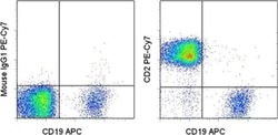
- Staining of normal human peripheral blood cells with Anti-Human CD19 APC (Product # 17-0199-42) and Mouse IgG1 K Isotype Control PE-Cyanine7 (Product # 25-4714-80) (left) or Anti-Human/Non-Human Primate CD2 PE-Cyanine7 (right). Cells in the lymphocyte gate were used for analysis.

Supportive validation
- Submitted by
- Invitrogen Antibodies (provider)
- Main image
- Experimental details
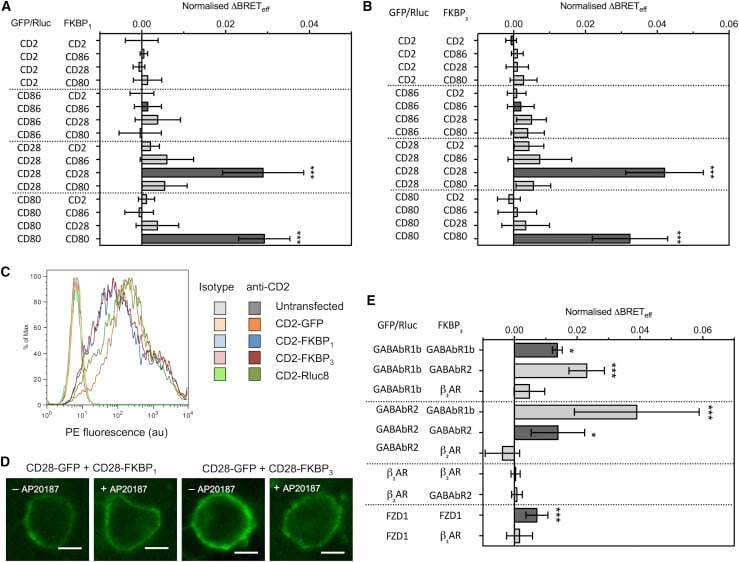
- Figure 2 Type-4 BRET assay applied to known controls. ( A ) The mean Delta BRET eff of type-1 transmembrane protein controls using the FKBP 1 type-4 BRET assay. Only the known dimers CD28 and CD80 exhibited significantly nonzero Delta BRET eff when expressed as homomeric pairs. Dark gray bars indicate like-like pairs. ( B ) The mean Delta BRET eff of type-1 transmembrane protein controls using the FKBP 3 type-4 BRET assay. Delta BRET eff is larger for CD28 and CD80 than with the FKBP 1 approach. ( C ) A histogram of CD2 expression in HEK-293T cells transfected with 1 mu g of differently tagged forms of CD2, as measured by flow cytometry. ( D ) Confocal microscopy of CD28-GFP coexpressed with CD28-FKBP 1/3 in the presence and absence of AP20187. No clear GFP internalization was evident. ( E ) The mean Delta BRET eff of GPCRs of known stoichiometry using the FKBP 3 type-4 BRET assay. For all panels, bars indicate mean +- SD. Probability is indicated for difference from Delta BRET eff = 0; * p < 0.05, *** p < 0.005. All data are the result of n >= 3 independent experiments.