Explore
 Validate
 Learn
Learn Flow cytometry
Antibody data
- Antibody Data
- Antigen structure
- References [6]
- Comments [0]
- Validations
- Flow cytometry [1]
- Other assay [1]
Submit
Validation data
Reference
Comment
Report error
- Product number
- 48-0029-42 - Provider product page

- Provider
- Invitrogen Antibodies
- Product name
- CD2 Monoclonal Antibody (RPA-2.10), eFluor™ 450, eBioscience™
- Antibody type
- Monoclonal
- Antigen
- Other
- Description
- Description: The RPA-2.10 monoclonal antibody reacts with human CD2, a 50 kDa cell surface receptor expressed by a majority of thymocytes, all mature T cells and subset of NK cells. CD2 is a ligand for CD58 in the human and is involved in adhesion and activation of T cells. RPA-2.10 blocks mixed lymphocyte reaction.
- Antibody clone number
- RPA-2.10
- Concentration
- 5 µL/Test
Submitted references Constraints on GPCR Heterodimerization Revealed by the Type-4 Induced-Association BRET Assay.
Tissue factor expression by myeloid cells contributes to protective immune response against Mycobacterium tuberculosis infection.
Monocyte-activation phenotypes are associated with biomarkers of inflammation and coagulation in chronic HIV infection.
Evidence for innate immune system activation in HIV type 1-infected elite controllers.
Decreases in colonic and systemic inflammation in chronic HIV infection after IL-7 administration.
A monoclonal antibody selection for immunohistochemical examination of lymphoid tissues from non-human primates.
Felce JH, MacRae A, Davis SJ
Biophysical journal 2019 Jan 8;116(1):31-41
Biophysical journal 2019 Jan 8;116(1):31-41
Tissue factor expression by myeloid cells contributes to protective immune response against Mycobacterium tuberculosis infection.
Venkatasubramanian S, Tripathi D, Tucker T, Paidipally P, Cheekatla S, Welch E, Raghunath A, Jeffers A, Tvinnereim AR, Schechter ME, Andrade BB, Mackman N, Idell S, Vankayalapati R
European journal of immunology 2016 Feb;46(2):464-79
European journal of immunology 2016 Feb;46(2):464-79
Monocyte-activation phenotypes are associated with biomarkers of inflammation and coagulation in chronic HIV infection.
Wilson EM, Singh A, Hullsiek KH, Gibson D, Henry WK, Lichtenstein K, Önen NF, Kojic E, Patel P, Brooks JT, Sereti I, Baker JV, Study to Understand the Natural History of HIV/AIDS in the Era of Effective Therapy (SUN Study) Investigators
The Journal of infectious diseases 2014 Nov 1;210(9):1396-406
The Journal of infectious diseases 2014 Nov 1;210(9):1396-406
Evidence for innate immune system activation in HIV type 1-infected elite controllers.
Krishnan S, Wilson EM, Sheikh V, Rupert A, Mendoza D, Yang J, Lempicki R, Migueles SA, Sereti I
The Journal of infectious diseases 2014 Mar;209(6):931-9
The Journal of infectious diseases 2014 Mar;209(6):931-9
Decreases in colonic and systemic inflammation in chronic HIV infection after IL-7 administration.
Sereti I, Estes JD, Thompson WL, Morcock DR, Fischl MA, Croughs T, Beq S, Lafaye de Micheaux S, Yao MD, Ober A, Wilson EM, Natarajan V, Imamichi H, Boulassel MR, Lederman MM, Routy JP
PLoS pathogens 2014 Jan;10(1):e1003890
PLoS pathogens 2014 Jan;10(1):e1003890
A monoclonal antibody selection for immunohistochemical examination of lymphoid tissues from non-human primates.
Kap YS, van Meurs M, van Driel N, Koopman G, Melief MJ, Brok HP, Laman JD, 't Hart BA
The journal of histochemistry and cytochemistry : official journal of the Histochemistry Society 2009 Dec;57(12):1159-67
The journal of histochemistry and cytochemistry : official journal of the Histochemistry Society 2009 Dec;57(12):1159-67
No comments: Submit comment
Supportive validation
- Submitted by
- Invitrogen Antibodies (provider)
- Main image

- Experimental details
- Staining of normal human peripheral blood cells with Anti-Human CD19 FITC (Product # 11-0199-42) and Mouse IgG1 K Isotype Control eFluor® 450 (Product # 48-4714-82) (left) or Anti-Human/Non-Human Primate CD2 eFluor® 450 (right). Cells in the lymphocyte gate were used for analysis.

Supportive validation
- Submitted by
- Invitrogen Antibodies (provider)
- Main image
- Experimental details
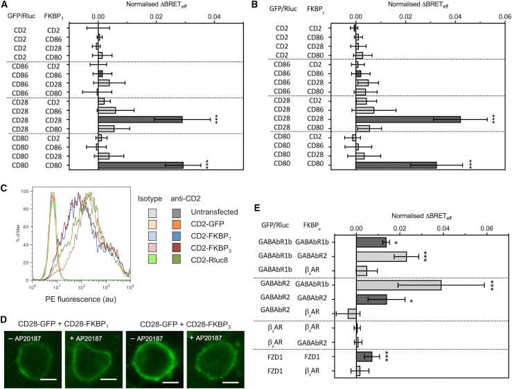
- Figure 2 Type-4 BRET assay applied to known controls. ( A ) The mean Delta BRET eff of type-1 transmembrane protein controls using the FKBP 1 type-4 BRET assay. Only the known dimers CD28 and CD80 exhibited significantly nonzero Delta BRET eff when expressed as homomeric pairs. Dark gray bars indicate like-like pairs. ( B ) The mean Delta BRET eff of type-1 transmembrane protein controls using the FKBP 3 type-4 BRET assay. Delta BRET eff is larger for CD28 and CD80 than with the FKBP 1 approach. ( C ) A histogram of CD2 expression in HEK-293T cells transfected with 1 mu g of differently tagged forms of CD2, as measured by flow cytometry. ( D ) Confocal microscopy of CD28-GFP coexpressed with CD28-FKBP 1/3 in the presence and absence of AP20187. No clear GFP internalization was evident. ( E ) The mean Delta BRET eff of GPCRs of known stoichiometry using the FKBP 3 type-4 BRET assay. For all panels, bars indicate mean +- SD. Probability is indicated for difference from Delta BRET eff = 0; * p < 0.05, *** p < 0.005. All data are the result of n >= 3 independent experiments.