Explore
 Validate
 Learn
Learn Flow cytometry
Antibody data
- Antibody Data
- Antigen structure
- References [1]
- Comments [0]
- Validations
- Flow cytometry [2]
- Other assay [1]
Submit
Validation data
Reference
Comment
Report error
- Product number
- 62-0029-42 - Provider product page

- Provider
- Invitrogen Antibodies
- Product name
- CD2 Monoclonal Antibody (RPA-2.10), Super Bright™ 436, eBioscience™
- Antibody type
- Monoclonal
- Antigen
- Other
- Description
- Description: The RPA-2.10 monoclonal antibody reacts with human CD2, a 50 kDa cell surface receptor expressed by a majority of thymocytes, all mature T cells and subset of NK cells. CD2 is a ligand for CD58 in the human and is involved in adhesion and activation of T cells. RPA-2.10 blocks mixed lymphocyte reaction. RPA-2.10 crossreacts to non-human primates and pigs. Applications Reported: This RPA-2.10 antibody has been reported for use in flow cytometric analysis. Applications Tested: This RPA-2.10 antibody has been pre-diluted and tested by flow cytometric analysis. This may be used at 5 µL (0.125 µg) per test. A test is defined as the amount (µg) of antibody that will stain a cell sample in a final volume of 100 µL. Cell number should be determined empirically but can range from 10^5 to 10^8 cells/test. Super Bright 436 can be excited with the violet laser line (405 nm) and emits at 436 nm. We recommend using a 450/50 bandpass filter, or equivalent. Please make sure that your instrument is capable of detecting this fluorochrome. When using two or more Super Bright dye-conjugated antibodies in a staining panel, it is recommended to use Super Bright Complete Staining Buffer (Product # SB-4401) to minimize any non-specific polymer interactions. Please refer to the datasheet for Super Bright Staining Buffer for more information. Excitation: 405 nm; Emission: 436 nm; Laser: Violet Laser Super Bright Polymer Dyes are sold under license from Becton, Dickinson and Company.
- Reactivity
- Human, Porcine
- Host
- Mouse
- Isotype
- IgG
- Antibody clone number
- RPA-2.10
- Vial size
- 100 Tests
- Concentration
- 5 µL/Test
- Storage
- 4° C, store in dark, DO NOT FREEZE!
Submitted references Constraints on GPCR Heterodimerization Revealed by the Type-4 Induced-Association BRET Assay.
Felce JH, MacRae A, Davis SJ
Biophysical journal 2019 Jan 8;116(1):31-41
Biophysical journal 2019 Jan 8;116(1):31-41
No comments: Submit comment
Supportive validation
- Submitted by
- Invitrogen Antibodies (provider)
- Main image

- Experimental details
- Normal human peripheral blood cells were stained with CD19 Monoclonal Antibody, APC (Product # 17-0199) and Mouse IgG1 kappa Isotype Control, Super Bright 436 (Product # 62-4714) (left) or CD2 Monoclonal Antibody, Super Bright 436 (right). Cells in the lymphocyte gate were used for analysis.

Supportive validation
- Submitted by
- Invitrogen Antibodies (provider)
- Main image
- Experimental details
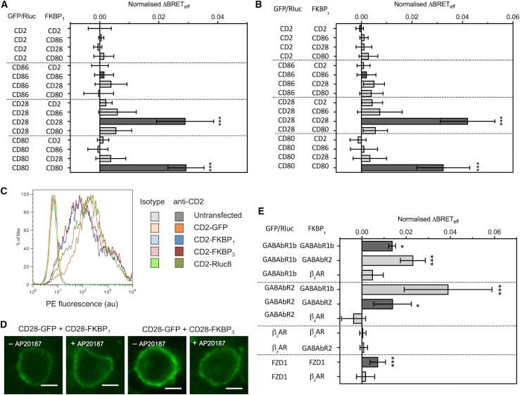
- Figure 2 Type-4 BRET assay applied to known controls. ( A ) The mean Delta BRET eff of type-1 transmembrane protein controls using the FKBP 1 type-4 BRET assay. Only the known dimers CD28 and CD80 exhibited significantly nonzero Delta BRET eff when expressed as homomeric pairs. Dark gray bars indicate like-like pairs. ( B ) The mean Delta BRET eff of type-1 transmembrane protein controls using the FKBP 3 type-4 BRET assay. Delta BRET eff is larger for CD28 and CD80 than with the FKBP 1 approach. ( C ) A histogram of CD2 expression in HEK-293T cells transfected with 1 mu g of differently tagged forms of CD2, as measured by flow cytometry. ( D ) Confocal microscopy of CD28-GFP coexpressed with CD28-FKBP 1/3 in the presence and absence of AP20187. No clear GFP internalization was evident. ( E ) The mean Delta BRET eff of GPCRs of known stoichiometry using the FKBP 3 type-4 BRET assay. For all panels, bars indicate mean +- SD. Probability is indicated for difference from Delta BRET eff = 0; * p < 0.05, *** p < 0.005. All data are the result of n >= 3 independent experiments.