Explore
 Validate
 Learn
LearnUM500006CF
antibody from Invitrogen Antibodies
Targeting: CD2
SRBC
 Western blot Immunocytochemistry Immunohistochemistry Flow cytometry
 Chromatin Immunoprecipitation Other assay
Antibody data
- Antibody Data
- Antigen structure
- References [0]
- Comments [0]
- Validations
- Western blot [4]
- Immunocytochemistry [1]
- Immunohistochemistry [7]
- Flow cytometry [1]
- Other assay [1]
Submit
Validation data
Reference
Comment
Report error
- Product number
- UM500006CF - Provider product page

- Provider
- Invitrogen Antibodies
- Product name
- CD2 Monoclonal Antibody (UMAB6), UltraMAB™
- Antibody type
- Monoclonal
- Antigen
- Other
- Reactivity
- Human
- Host
- Mouse
- Isotype
- IgG
- Antibody clone number
- UMAB6
- Vial size
- 100 µg
- Concentration
- 1 mg/mL
- Storage
- -20° C, Avoid Freeze/Thaw Cycles
No comments: Submit comment
Supportive validation
- Submitted by
- Invitrogen Antibodies (provider)
- Main image

- Experimental details
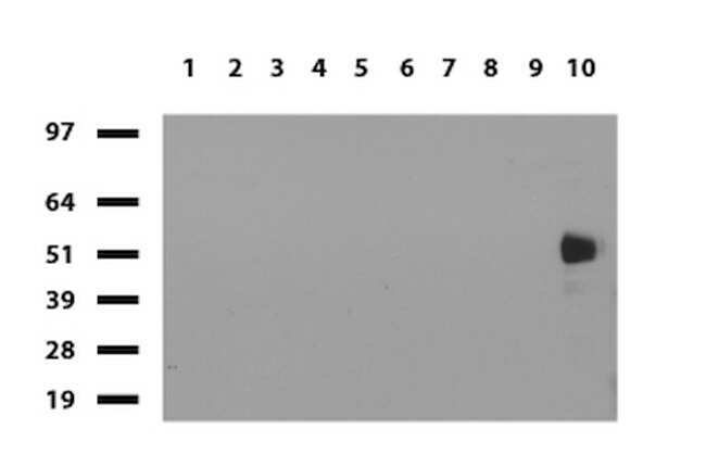
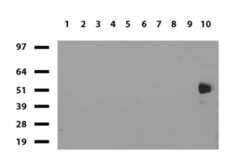
- Western blot of human tissue lysates (15 µg) from 10 different tissues (1: Testis, 2: Omentum, 3: Uterus, 4: Breast, 5: Brain, 6: Liver, 7: Ovary, 8: Thyroid 9: Colon, 10: Spleen). Dilution: 1:500.
- Submitted by
- Invitrogen Antibodies (provider)
- Main image
- Experimental details
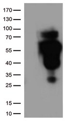
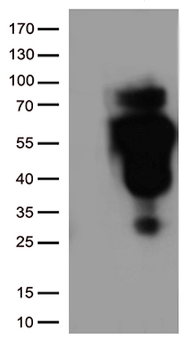
- HEK293T cells were transfected with the pCMV6-ENTRY control (Left lane) or pCMV6-ENTRY CD2(RC206612, Right lane) cDNA for 48 hrs and lysed. Equivalent amounts of cell lysates (5 µg per lane) were separated by SDS-PAGE and immunoblotted with anti-CD2. (1:500)
- Submitted by
- Invitrogen Antibodies (provider)
- Main image
- Experimental details
- Western blot of human tissue lysates (15 µg) from 10 different tissues (1: Testis, 2: Omentum, 3: Uterus, 4: Breast, 5: Brain, 6: Liver, 7: Ovary, 8: Thyroid 9: Colon, 10: Spleen). Dilution: 1:500.

- Submitted by
- Invitrogen Antibodies (provider)
- Main image
- Experimental details
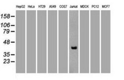
- Western blot analysis of extracts (35 µg) from 9 different cell lines by using anti-CD2 monoclonal antibody (Clone UMAB6). (1:1000)

Supportive validation
- Submitted by
- Invitrogen Antibodies (provider)
- Main image
- Experimental details
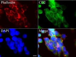
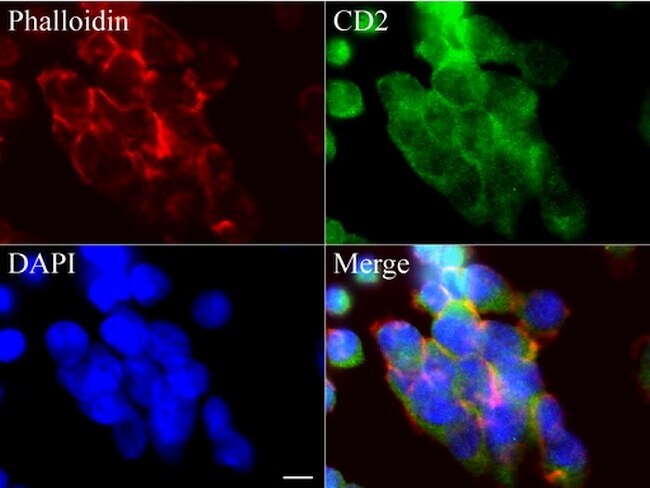
- Immunofluorescent staining of Jurkat cells using anti-CD2 mouse monoclonal antibody (UM500006, green, 1:100). Actin filaments were labeled with Alexa Fluor 594 Phalloidin (red), and nuclear with DAPI (blue). Scale bar, 8µm.
Supportive validation
- Submitted by
- Invitrogen Antibodies (provider)
- Main image
- Experimental details
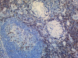
- IHC staining of paraffin-embedded human tonsil using anti-CD2 clone UMAB6 mouse monoclonal antibody at 1:200 of 0.6mg/mL and detection with Polink2 Broad HRP DAB. UM500006 requires heat-induced epitope retrieval with Accel pH8.7 at 95-100C 30 minutes [do not let boil] or 10 min in pressure cooker. The image shows strong membranous and cytoplasmic staining in >50 % of germinal center cells of tonsil and
- Submitted by
- Invitrogen Antibodies (provider)
- Main image
- Experimental details
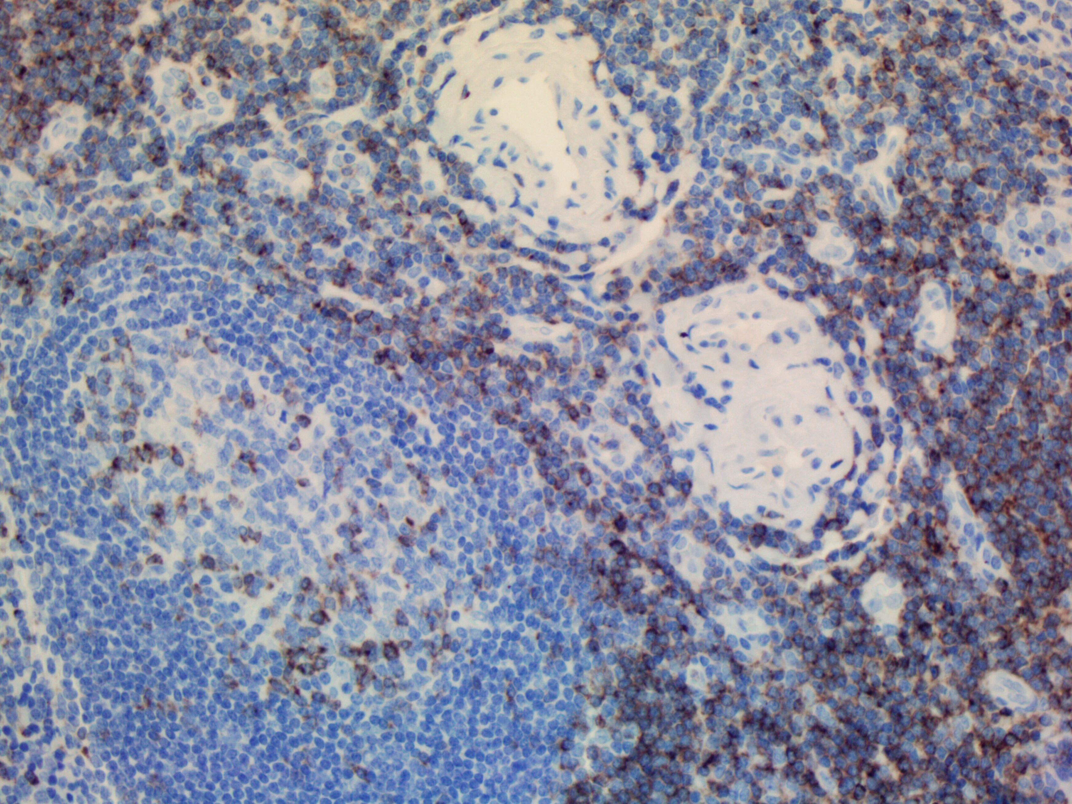
- IHC staining of FFPE human spleen using anti-CD2 clone UMAB6 mouse monoclonal antibody at 1:200 and detection with Polink2 Broad HRP DAB. UM500006 requires heat-induced epitope retrieval with Citrate pH6.O. The image shows strong membranous and cytoplasmic staining.

- Submitted by
- Invitrogen Antibodies (provider)
- Main image
- Experimental details
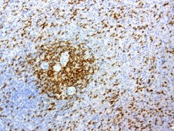
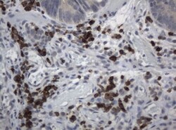
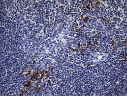
- Immunohistochemical staining of paraffin-embedded human lymphoma tissue using anti-CD2 mouse monoclonal antibody. (Clone UMAB6, dilution 1:100; heat-induced epitope retrieval by 10mM citric buffer, pH6.0, 120°C for 3min)

- Submitted by
- Invitrogen Antibodies (provider)
- Main image
- Experimental details
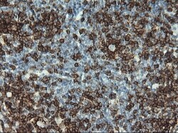
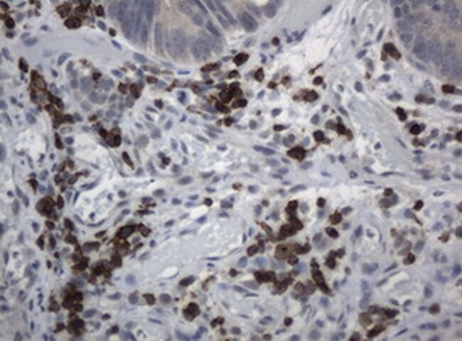
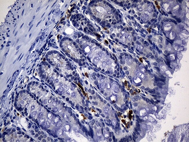
- Immunohistochemical staining of paraffin-embedded Adenocarcinoma of colon tissue using anti-CD2 mouse monoclonal antibody. (Clone UMAB6, dilution 1:100; heat-induced epitope retrieval by 10mM citric buffer, pH6.0, 120°C for 3min)
- Submitted by
- Invitrogen Antibodies (provider)
- Main image

- Experimental details
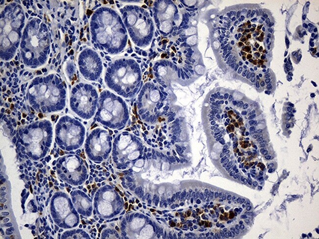
- Immunohistochemical staining of paraffin-embedded mouse ascending colon tissue anti-CD2 clone UMAB6 mouse monoclonal antibody. (Heat-induced epitope retrieval by 1mM EDTA in 10mM Tris buffer (pH8.5) at 120°C for 3 min, UM500006)(1:500)
- Submitted by
- Invitrogen Antibodies (provider)
- Main image
- Experimental details
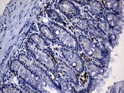
- Immunohistochemical staining of paraffin-embedded mouse colon tissue within the normal limits using anti-CD2 mouse monoclonal antibody. (Heat-induced epitope retrieval by 1mM EDTA in 10mM Tris buffer (pH8.5) at 120°C for 3 min, UM500006)(1:500)
- Submitted by
- Invitrogen Antibodies (provider)
- Main image
- Experimental details
- Immunohistochemical staining of paraffin-embedded mouse spleen tissue using anti-CD2 clone UMAB6 mouse monoclonal antibody. (Heat-induced epitope retrieval by 1mM EDTA in 10mM Tris buffer (pH8.5) at 120°C for 3 min, UM500006)(1:500).

Supportive validation
- Submitted by
- Invitrogen Antibodies (provider)
- Main image
- Experimental details
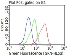
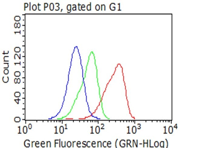
- Flow cytometric analysis of living Jurkat cells, using anti-CD2 antibody (UM500006, Red), compared to an isotype control (green), and a PBS control (blue). (1:100)
Supportive validation
- Submitted by
- Invitrogen Antibodies (provider)
- Main image
- Experimental details
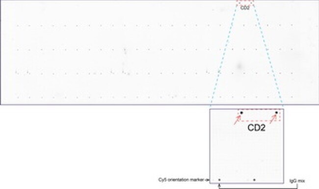
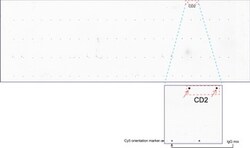
- OriGene overexpression protein microarray chip was immunostained with UltraMAB anti-CD2 mouse monoclonal antibody (Clone UMAB6). The positive reactive proteins are highlighted with two red arrows in the enlarged subarray. All the positive controls spotted in this subarray are also labeled for clarification. These data show that UltraMAB anti-CD2 (Clone UMAB6) very specifically recognizes Cd2 antigen on OriGene protein microarray chip.