Explore
 Validate
 Learn
Learn Western blot
 Immunoprecipitation
 Flow cytometry
Antibody data
- Antibody Data
- Antigen structure
- References [8]
- Comments [0]
- Validations
- Flow cytometry [7]
Submit
Validation data
Reference
Comment
Report error
- Product number
- MA0200 - Provider product page

- Provider
- Invitrogen Antibodies
- Product name
- CD2 Monoclonal Antibody (TS2/18)
- Antibody type
- Monoclonal
- Antigen
- Other
- Description
- MA0200 targets CD2 in FACS, IA, IP, and WB applications and shows reactivity with Human samples. This antibody does not react with rat tissue in Western blot applications. The MA0200 immunogen is human LFA-2 (CD2). MA0200 detects CD2 which has a predicted molecular weight of approximately 37 kDa. This product has been tested for endotoxins by limulus amoebocyte lysate (LAL) assay and contains an endotoxin concentration of less than or equal to 10 endotoxin units per milligram (EU/mg).
- Reactivity
- Human
- Host
- Mouse
- Isotype
- IgG
- Antibody clone number
- TS2/18
- Vial size
- 200 μg
- Concentration
- 1.0 mg/mL
- Storage
- -20°C
Submitted references Antibody blocking of MHC II on human activated regulatory T cells abrogates their suppressive potential.
Mechanisms of Cellular Avidity Regulation in CD2-CD58-Mediated T Cell Adhesion.
Different roles of the CD2 and LFA-1 T-cell co-receptors for regulating cytotoxic, proliferative, and cytokine responses of human V gamma 9/V delta 2 T cells.
Selective deletion of antigen-specific, activated T cells by a humanized MAB to CD2 (MEDI-507) is mediated by NK cells.
Selective deletion of antigen-specific, activated T cells by a humanized MAB to CD2 (MEDI-507) is mediated by NK cells.
Intercellular adhesion molecule-1 and leukocyte function-associated antigen-3 provide costimulation for superantigen-induced T lymphocyte proliferation in the absence of a specific presenting molecule.
CD28-mediated up-regulation of beta 1-integrin adhesion involves phosphatidylinositol 3-kinase.
The T lymphocyte glycoprotein CD2 binds the cell surface ligand LFA-3.
Peiser M, Becht A, Wanner R
Allergy 2007 Jul;62(7):773-80
Allergy 2007 Jul;62(7):773-80
Mechanisms of Cellular Avidity Regulation in CD2-CD58-Mediated T Cell Adhesion.
Zhu DM, Dustin ML, Cairo CW, Thatte HS, Golan DE
ACS chemical biology 2006 Nov 21;1(10):649-58
ACS chemical biology 2006 Nov 21;1(10):649-58
Different roles of the CD2 and LFA-1 T-cell co-receptors for regulating cytotoxic, proliferative, and cytokine responses of human V gamma 9/V delta 2 T cells.
Wang P, Malkovsky M
Molecular medicine (Cambridge, Mass.) 2000 Mar;6(3):196-207
Molecular medicine (Cambridge, Mass.) 2000 Mar;6(3):196-207
Selective deletion of antigen-specific, activated T cells by a humanized MAB to CD2 (MEDI-507) is mediated by NK cells.
Branco L, Barren P, Mao SY, Pfarr D, Kaplan R, Postema C, Langermann S, Koenig S, Johnson S
Transplantation 1999 Nov 27;68(10):1588-96
Transplantation 1999 Nov 27;68(10):1588-96
Selective deletion of antigen-specific, activated T cells by a humanized MAB to CD2 (MEDI-507) is mediated by NK cells.
Branco L, Barren P, Mao SY, Pfarr D, Kaplan R, Postema C, Langermann S, Koenig S, Johnson S
Transplantation 1999 Nov 27;68(10):1588-96
Transplantation 1999 Nov 27;68(10):1588-96
Intercellular adhesion molecule-1 and leukocyte function-associated antigen-3 provide costimulation for superantigen-induced T lymphocyte proliferation in the absence of a specific presenting molecule.
Lamphear JG, Stevens KR, Rich RR
Journal of immunology (Baltimore, Md. : 1950) 1998 Jan 15;160(2):615-23
Journal of immunology (Baltimore, Md. : 1950) 1998 Jan 15;160(2):615-23
CD28-mediated up-regulation of beta 1-integrin adhesion involves phosphatidylinositol 3-kinase.
Zell T, Hunt SW 3rd, Mobley JL, Finkelstein LD, Shimizu Y
Journal of immunology (Baltimore, Md. : 1950) 1996 Feb 1;156(3):883-6
Journal of immunology (Baltimore, Md. : 1950) 1996 Feb 1;156(3):883-6
The T lymphocyte glycoprotein CD2 binds the cell surface ligand LFA-3.
Selvaraj P, Plunkett ML, Dustin M, Sanders ME, Shaw S, Springer TA
Nature 1987 Mar 26-Apr 1;326(6111):400-3
Nature 1987 Mar 26-Apr 1;326(6111):400-3
No comments: Submit comment
Supportive validation
- Submitted by
- Invitrogen Antibodies (provider)
- Main image
- Experimental details
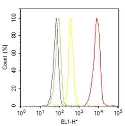
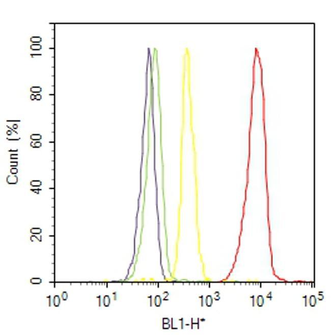
- Flow cytometry analysis of CD2 / LFA-2 was done on Jurkat cells. Cells were fixed with 70% ethanol for 10 minutes, permeabilized with 0.25% Triton™ X-100 for 20 minutes, and blocked with 5% BSA for 30 minutes at room temperature. Cells were labeled with CD2 / LFA-2 Mouse Monoclonal Antibody (MA0200, red histogram) or with mouse isotype control (yellow histogram) at 3-5 ug/million cells in 2.5% BSA. After incubation at room temperature for 2 hours, the cells were labeled with Alexa Fluor® 488 Rabbit Anti-Mouse Secondary Antibody (A11059) at a dilution of 1:400 for 30 minutes at room temperature. The representative 10,000 cells were acquired and analyzed for each sample using an Attune® Acoustic Focusing Cytometer. The purple histogram represents unstained control cells and the green histogram represents no-primary-antibody control.
- Submitted by
- Invitrogen Antibodies (provider)
- Main image
- Experimental details
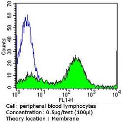
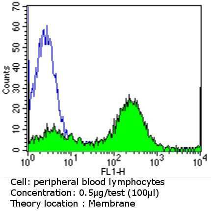
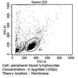
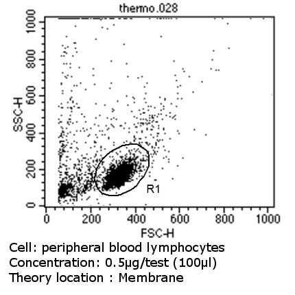
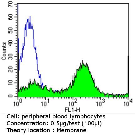
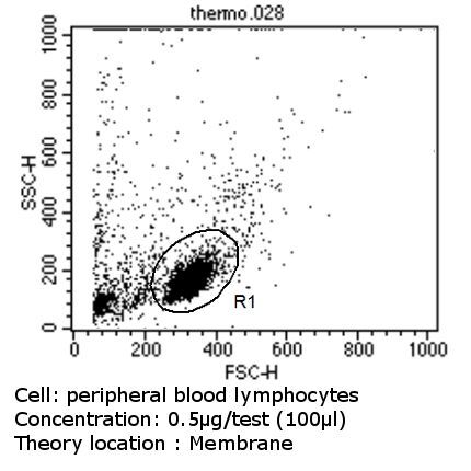
- Flow cytometry analysis of CD2 in PBMC cells (green) compared to an isotype control (blue). Human blood was collected, combined with a hydrophilic polysaccharide, centrifuged, transferred to a conical tube and washed with PBS. 50 µL of cell solution was added to each tube at a dilution of 2x10^7 cells/mL, followed by the addition of 50 µL of isotype control and primary antibody (Product # MA0200) at a dilution of 0.5 µg/test. Cells were incubated for 30 min at 4ºC and washed with a cell buffer, followed by incubation with a DyLight 488-conjugated goat anti-mouse IgG (H+L) secondary for 30 min at 4ºC in the dark. FACS analysis was performed using 400 µL of cell buffer.
- Submitted by
- Invitrogen Antibodies (provider)
- Main image
- Experimental details
- Flow cytometry analysis of CD2 in PBMC cells (green) compared to an isotype control (blue). Human blood was collected, combined with a hydrophilic polysaccharide, centrifuged, transferred to a conical tube and washed with PBS. 50 µL of cell solution was added to each tube at a dilution of 2x10^7 cells/mL, followed by the addition of 50 µL of isotype control and primary antibody (Product # MA0200) at a dilution of 0.5 µg/test. Cells were incubated for 30 min at 4ºC and washed with a cell buffer, followed by incubation with a DyLight 488-conjugated goat anti-mouse IgG (H+L) secondary for 30 min at 4ºC in the dark. FACS analysis was performed using 400 µL of cell buffer.
- Submitted by
- Invitrogen Antibodies (provider)
- Main image

- Experimental details
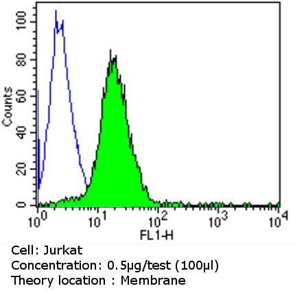
- Flow cytometry analysis of CD2 in Jurkat cells (green) compared to an isotype control (blue). Cells were harvested, adjusted to a concentration of 1-5x10^6 cells/mL, fixed with 2% paraformaldehyde and washed with PBS. Cells were blocked with a 2% solution of BSA-PBS for 30 min at room temperature and incubated with a CD2 monoclonal antibody (Product # MA0200) at a dilution of 0.5 µg/test for 60 min at room temperature. Cells were then incubated for 40 min at room temperature in the dark using a Dylight 488-conjugated goat anti-mouse IgG (H+L) secondary antibody and re-suspended in PBS for FACS analysis.

- Submitted by
- Invitrogen Antibodies (provider)
- Main image

- Experimental details
- Flow cytometry analysis of CD2 in PBMC cells (green) compared to an isotype control (blue). Human blood was collected, combined with a hydrophilic polysaccharide, centrifuged, transferred to a conical tube and washed with PBS. 50 µL of cell solution was added to each tube at a dilution of 2x10^7 cells/mL, followed by the addition of 50 µL of isotype control and primary antibody (Product # MA0200) at a dilution of 0.5 µg/test. Cells were incubated for 30 min at 4ºC and washed with a cell buffer, followed by incubation with a DyLight 488-conjugated goat anti-mouse IgG (H+L) secondary for 30 min at 4ºC in the dark. FACS analysis was performed using 400 µL of cell buffer.
- Submitted by
- Invitrogen Antibodies (provider)
- Main image

- Experimental details
- Flow cytometry analysis of CD2 in Jurkat cells (green) compared to an isotype control (blue). Cells were harvested, adjusted to a concentration of 1-5x10^6 cells/mL, fixed with 2% paraformaldehyde and washed with PBS. Cells were blocked with a 2% solution of BSA-PBS for 30 min at room temperature and incubated with a CD2 monoclonal antibody (Product # MA0200) at a dilution of 0.5 µg/test for 60 min at room temperature. Cells were then incubated for 40 min at room temperature in the dark using a Dylight 488-conjugated goat anti-mouse IgG (H+L) secondary antibody and re-suspended in PBS for FACS analysis.