 Explore
 Validate
 Learn
Learn Western blot
Antibody data
- Antibody Data
- Antigen structure
- References [4]
- Comments [0]
- Validations
- Western blot [1]
- Immunohistochemistry [1]
- Flow cytometry [2]
Submit
Validation data
Reference
Comment
Report error
- Product number
- MA5-11373 - Provider product page

- Provider
- Invitrogen Antibodies
- Product name
- CD2 Monoclonal Antibody (AB75)
- Antibody type
- Monoclonal
- Antigen
- Recombinant protein fragment
- Description
- MA5-11373 targets CD2 in IHC (P) and FACS applications and shows reactivity with Human samples. The MA5-11373 immunogen is recombinant fragment encoding the external domain of the human CD2 protein.
- Reactivity
- Human
- Host
- Mouse
- Isotype
- IgG
- Antibody clone number
- AB75
- Vial size
- 500 μL
- Concentration
- Conc. Not Determined
- Storage
- 4°C
Submitted references Nontuberculous mycobacterial infection with concurrent IgG4-related lymphadenopathy.
Infectious mononucleosis mimicking malignant T-cell lymphoma in the nasopharynx: a case report and review of the literature.
Human Immunodeficiency Virus (HIV)-Negative and Human Herpes Virus-8 (HHV-8)-Positive Primary Effusion Lymphoma: A Case Report and Review of the Literature.
Anaplastic large cell lymphoma with paraneoplastic leukocytosis: a clinicopathological analysis of five cases.
Liu TT, Weng SW, Wang MC, Huang WT
APMIS : acta pathologica, microbiologica, et immunologica Scandinavica 2016 Mar;124(3):216-20
APMIS : acta pathologica, microbiologica, et immunologica Scandinavica 2016 Mar;124(3):216-20
Infectious mononucleosis mimicking malignant T-cell lymphoma in the nasopharynx: a case report and review of the literature.
He HL, Wang MC, Huang WT
International journal of clinical and experimental pathology 2013;6(1):105-9
International journal of clinical and experimental pathology 2013;6(1):105-9
Human Immunodeficiency Virus (HIV)-Negative and Human Herpes Virus-8 (HHV-8)-Positive Primary Effusion Lymphoma: A Case Report and Review of the Literature.
Güven Karataş S, Bayrak R, Sahin Balçık O, Yalçın KS, Atıcı E, Akyıldız U, Koşar A
Turkish journal of haematology : official journal of Turkish Society of Haematology 2013 Mar;30(1):67-71
Turkish journal of haematology : official journal of Turkish Society of Haematology 2013 Mar;30(1):67-71
Anaplastic large cell lymphoma with paraneoplastic leukocytosis: a clinicopathological analysis of five cases.
Chang IW, Chen HK, Ma MC, Huang WT
APMIS : acta pathologica, microbiologica, et immunologica Scandinavica 2011 Nov;119(11):794-801
APMIS : acta pathologica, microbiologica, et immunologica Scandinavica 2011 Nov;119(11):794-801
No comments: Submit comment
Supportive validation
- Submitted by
- Invitrogen Antibodies (provider)
- Main image
- Experimental details
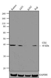
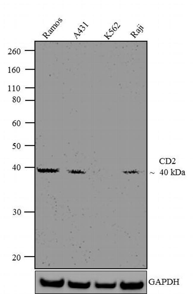
- Western blot analysis was performed on whole cell extracts (30 µg lysate) of Ramos (Lane1), A-431 (Lane2), K562 (Lane 3) and Raji (Lane4). The blots were probed with Anti-CD2 Mouse Monoclonal Antibody (Product # MA0200, 1:100-1:500 dilution) and detected by chemiluminescence using Goat anti-Mouse IgG (H+L) Secondary Antibody, HRP conjugate (Product # 62-6520, 1:4000 dilution). A 40 kDa band corresponding to CD2 was observed across cell lines tested expect for K562. Known quantity of protein samples were electrophoresed using Novex® NuPAGE® 12 % Bis-Tris gel (Product # NP0341BOX), XCell SureLock™ Electrophoresis System (Product # EI0002), and Novex® Sharp Pre-Stained Protein Standard (Product # LC5800). Resolved proteins were then transferred onto a nitrocellulose membrane with iBlot® 2 Dry Blotting System (Product # IB21001). The membrane was probed with the relevant primary and secondary Antibody using iBind™ Flex Western Starter Kit (Product # SLF2000S). Chemiluminescent detection was performed using Pierce™ ECL Western Blotting Substrate (Product # 32106).
Supportive validation
- Submitted by
- Invitrogen Antibodies (provider)
- Main image
- Experimental details
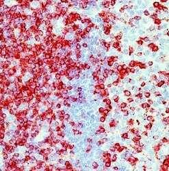
- Formalin-fixed, paraffin-embedded human tonsil stained with CD2 antibody using peroxidase-conjugate and AEC chromogen. Note membrane staining of T lymphocytes.

Supportive validation
- Submitted by
- Invitrogen Antibodies (provider)
- Main image

- Experimental details
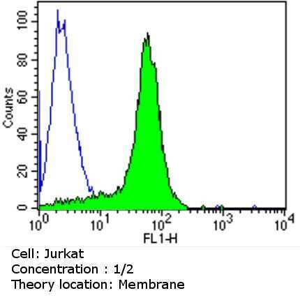
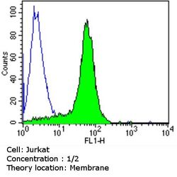
- Flow cytometry analysis of CD2 in Jurkat cells (green) compared to an isotype control (blue). Cells were harvested, adjusted to a concentration of 1-5x10^6 cells/mL, fixed with 2% paraformaldehyde and washed with PBS. Cells were blocked with a 2% solution of BSA-PBS for 30 min at room temperature and incubated with a CD2 monoclonal antibody (Product # MA5-11373) at a dilution of 2 µg/test for 60 min at room temperature. Cells were then incubated for 40 min at room temperature in the dark using a Dylight 488-conjugated goat anti-mouse IgG (H+L) secondary antibody and re-suspended in PBS for FACS analysis.
- Submitted by
- Invitrogen Antibodies (provider)
- Main image
- Experimental details
- Flow cytometry analysis of CD2 in Jurkat cells (green) compared to an isotype control (blue). Cells were harvested, adjusted to a concentration of 1-5x10^6 cells/mL, fixed with 2% paraformaldehyde and washed with PBS. Cells were blocked with a 2% solution of BSA-PBS for 30 min at room temperature and incubated with a CD2 monoclonal antibody (Product # MA5-11373) at a dilution of 2 µg/test for 60 min at room temperature. Cells were then incubated for 40 min at room temperature in the dark using a Dylight 488-conjugated goat anti-mouse IgG (H+L) secondary antibody and re-suspended in PBS for FACS analysis.
