Explore
 Validate
 Learn
Learn Flow cytometry
Antibody data
- Antibody Data
- Antigen structure
- References [0]
- Comments [0]
- Validations
- Flow cytometry [2]
Submit
Validation data
Reference
Comment
Report error
- Product number
- H006T03Y07 - Provider product page

- Provider
- Invitrogen Antibodies
- Product name
- CD16 Monoclonal Antibody (3G8), NovaFluor™ Yellow 730, eBioscience™
- Antibody type
- Monoclonal
- Antigen
- Other
- Description
- Description: This 3G8 monoclonal antibody reacts with human and non-human primate CD16, which is also known as the low-affinity Fc gamma RIII. CD16 exists as two distinct isoforms, Fc gamma RIIIA and Fc gamma RIIIB. In humans, Fc gamma RIIIA is expressed as a polypeptide-anchored form on monocytes, macrophages, and lymphocytes such as NK cells. T and B cells do not express this Fc receptor. Fc gamma RIIIB is also detected on neutrophils as a GPI-anchored form. Expression of CD16 on lymphocytes and monocytes is similar in non-human primates. However, while CD16 is not found on neutrophils in macaques and baboons, this receptor is detected on these cells in sooty mangabeys. Binding of IgG leads to activation of signal transduction pathways, resulting in antibody-dependent cell-mediated cytotoxicity (ADCC), phagocytosis, cytokine release, and antigen presentation.
- Antibody clone number
- 3G8
- Concentration
- 4 µL/Test
No comments: Submit comment
Supportive validation
- Submitted by
- Invitrogen Antibodies (provider)
- Main image

- Experimental details
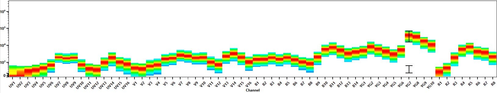
- Spectral signature for NovaFluor Yellow 730 collected on a 5-laser Cytek Aurora Full Spectrum flow cytometer using Cytek assay settings. Human peripheral blood mononuclear cells were stained with anti-human CD4 (SK3) and signatures displayed following gating on the lymphocyte population.
- Submitted by
- Invitrogen Antibodies (provider)
- Main image
- Experimental details
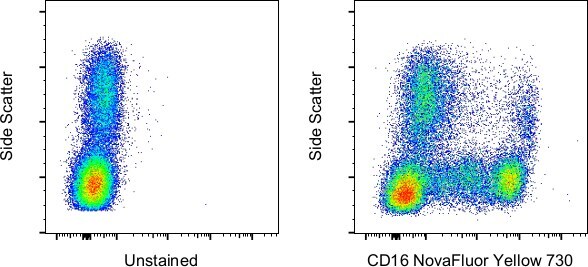
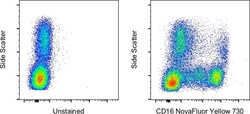
- Staining of normal human PBMCs with side scatter and unstained (left) or CD16 Monoclonal Antibody, NovaFluor Yellow 730 (right). Data was acquired in the YG8 channel on a 5-laser Cytek Aurora and singlet cells were used for analysis.