Explore
 Validate
 Learn
Learn Flow cytometry
Antibody data
- Antibody Data
- Antigen structure
- References [0]
- Comments [0]
- Validations
- Flow cytometry [9]
Submit
Validation data
Reference
Comment
Report error
- Product number
- MA5-44149 - Provider product page

- Provider
- Invitrogen Antibodies
- Product name
- CD16 Monoclonal Antibody (3G8), Alexa Fluor™ 700
- Antibody type
- Monoclonal
- Antigen
- Other
- Description
- This antibody detects both 158V and 158F allotype on natural NK cells.
- Reactivity
- Human
- Host
- Mouse
- Conjugate
- Near infrared dye
- Isotype
- IgG
- Antibody clone number
- 3G8
- Vial size
- 100 Tests
- Concentration
- 0.26 mg/mL
- Storage
- 4°C, store in dark, DO NOT FREEZE!
No comments: Submit comment
Supportive validation
- Submitted by
- Invitrogen Antibodies (provider)
- Main image
- Experimental details
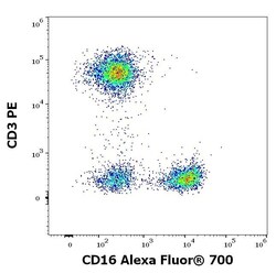
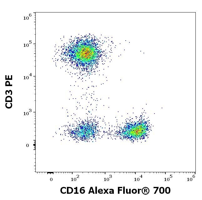
- Flow cytometry multicolor surface staining of human lymphocytes stained using anti-human CD16 (3G8) Alexa Fluor® 700 Monoclonal antibody (Product # MA5-44149), 4 µL reagent / 100 µL of peripheral whole blood) and anti-human CD3 (UCHT1) PE using a dilution of 20 µL reagent/100 µL of peripheral whole blood.
- Submitted by
- Invitrogen Antibodies (provider)
- Main image
- Experimental details
- Flow Cytometry of CD16 in human peripheral whole blood using CD16 Monoclonal Antibody, Alexa Fluor 700 (Product # MA5-44149) at a dilution of 4 µL/100 µL of peripheral whole blood.
- Conjugate
- Near infrared dye
- Submitted by
- Invitrogen Antibodies (provider)
- Main image
- Experimental details
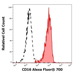
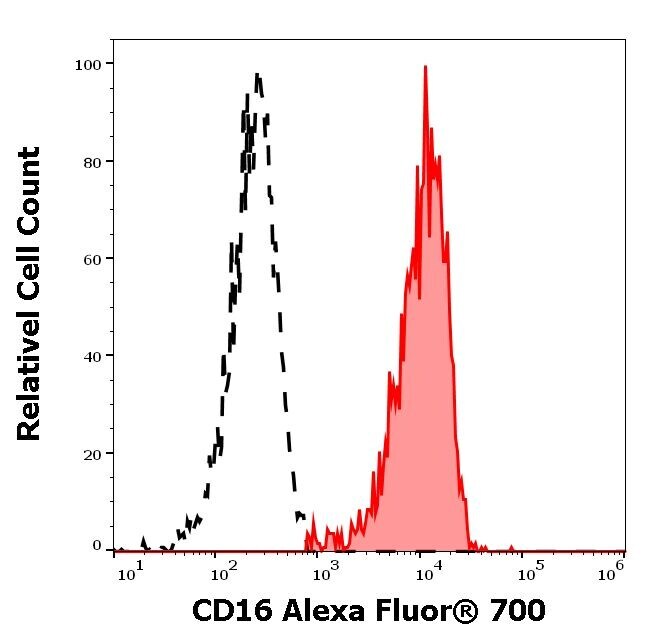
- Separation of human CD16 positive CD3 negative lymphocytes (red-filled) from CD16 negative CD3 positive lymphocytes (black-dashed) in flow cytometry analysis (surface staining) of human peripheral whole blood stained using anti-human CD16 (3G8) Alexa Fluor® 700 Monoclonal antibody (Product # MA5-44149) using a dilution of 4 µL reagent/100 µL of peripheral whole blood.
- Conjugate
- Near infrared dye
- Submitted by
- Invitrogen Antibodies (provider)
- Main image
- Experimental details
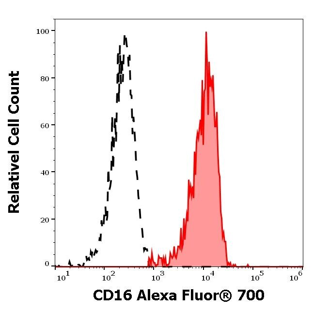
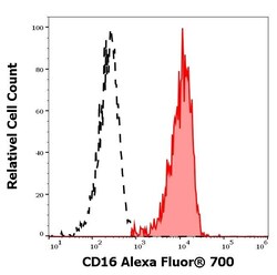
- Separation of human CD16 positive CD3 negative lymphocytes (red-filled) from CD16 negative CD3 positive lymphocytes (black-dashed) in flow cytometry analysis (surface staining) of human peripheral whole blood stained using anti-human CD16 (3G8) Alexa Fluor® 700 Monoclonal antibody (Product # MA5-44149) using a dilution of 4 µL reagent/100 µL of peripheral whole blood.
- Submitted by
- Invitrogen Antibodies (provider)
- Main image
- Experimental details
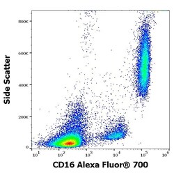
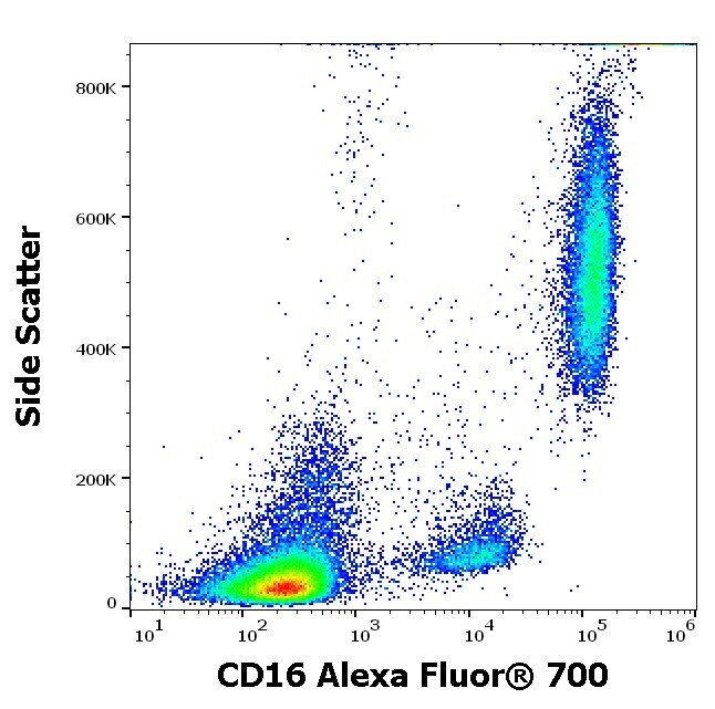
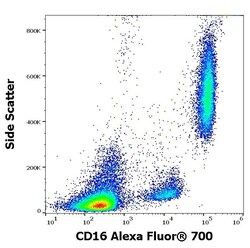
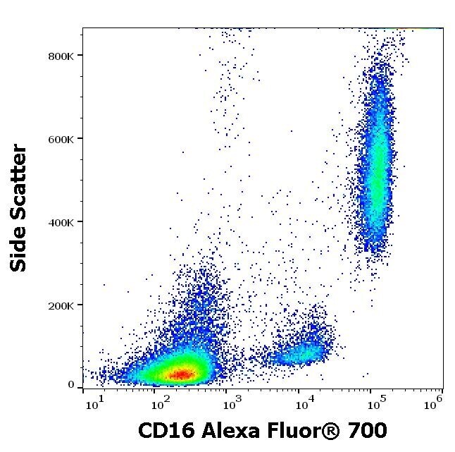
- Flow cytometry surface staining pattern of human peripheral whole blood stained using anti-human CD16 (3G8) Alexa Fluor® 700 Monoclonal antibody (Product # MA5-44149) using a dilution of 4 µL reagent/100 µL of peripheral whole blood.
- Submitted by
- Invitrogen Antibodies (provider)
- Main image
- Experimental details
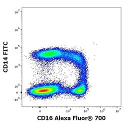
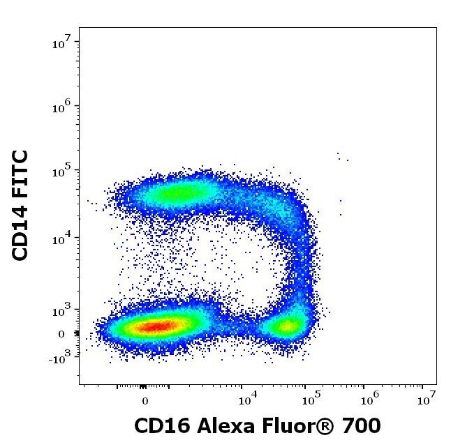
- Flow Cytometry analysis of CD16 using CD16 Monoclonal Antibody (3G8), Alexa Fluor™ 700 (Product # MA5-44149). Analysis of the antibody staining profile was performed on blood leukocytes isolated from peripheral whole blood by bulk erythrocyte lysis. Mouse monoclonal anti-human CD16 Alexa Fluor™ 700 antibody (clone 3G8) was used in concentration 10 µg/mL in stained stained blood sample (2 x 10⁶ cells) and Mouse monoclonal anti-human CD14 FITC antibody in concentration 7 µg/mL, respectively.
- Submitted by
- Invitrogen Antibodies (provider)
- Main image
- Experimental details
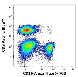
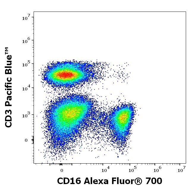
- Flow Cytometry analysis of CD16 using CD16 Monoclonal Antibody (3G8), Alexa Fluor™ 700 (Product # MA5-44149). Analysis of the antibody staining profile was performed on blood leukocytes isolated from peripheral whole blood by bulk erythrocyte lysis. Mouse monoclonal anti-human CD16 Alexa Fluor™ 700 antibody (clone 3G8) was used in concentration 10 µg/mL in stained stained blood sample (2 x 10⁶ cells) and Mouse monoclonal anti-human CD3 Pacific Blue™ antibody in concentration 9 µg/mL, respectively.
- Submitted by
- Invitrogen Antibodies (provider)
- Main image
- Experimental details
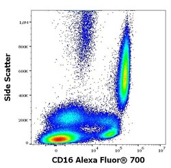
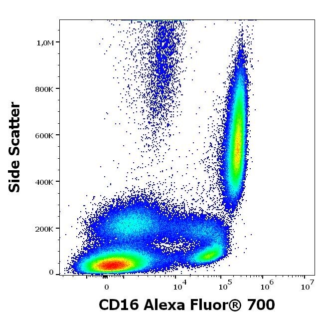
- Flow Cytometry analysis of CD16 using CD16 Monoclonal Antibody (3G8), Alexa Fluor™ 700 (Product # MA5-44149) . Analysis of the antibody staining profile was performed on blood leukocytes isolated from peripheral whole blood by bulk erythrocyte lysis. Mouse monoclonal anti-human CD16 Alexa Fluor™ 700 antibody (clone 3G8) was used in concentration 10 µg/mL in stained blood sample (2 x 10⁶ cells).
- Submitted by
- Invitrogen Antibodies (provider)
- Main image
- Experimental details
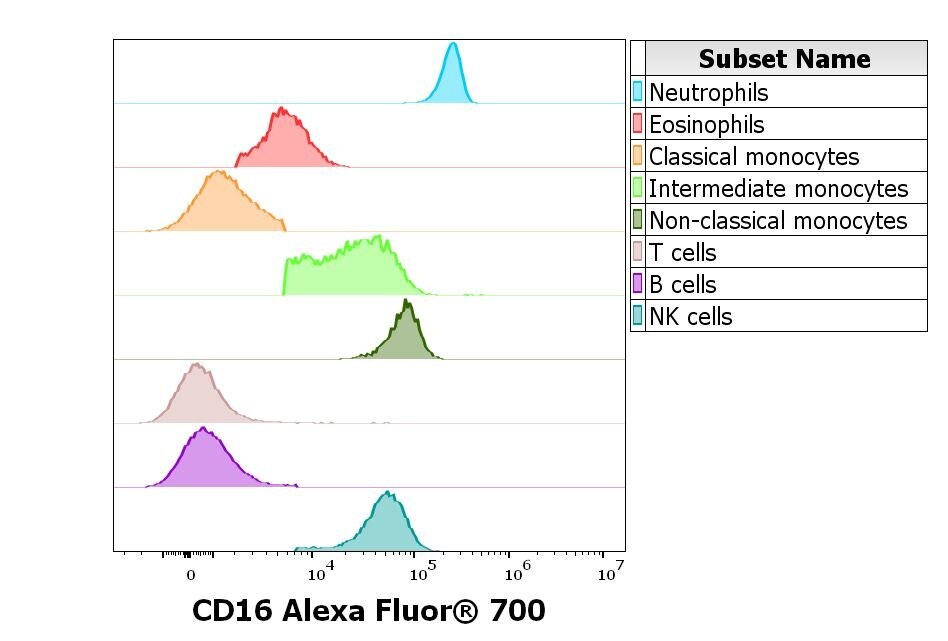
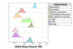
- Flow Cytometry analysis of CD16 using CD16 Monoclonal Antibody (3G8), Alexa Fluor™ 700 (Product # MA5-44149). Analysis of the antibody staining profile was performed on blood leukocytes isolated from peripheral whole blood by bulk erythrocyte lysis. Suspension of blood leukocytes (2 x 10⁶ cells) was added to the mixture of CD16 Alexa Fluor™ 700 antibody (clone 3G8, 10 µg/mL in stained blood sample), backbone antibody conjugates and monocyte blocking buffer, vortexed and incubated for 20 min. Stained sample was fixed with 2 mL of 10× diluted lysing solution for 10 min. Finally, samples were centrifuged (670 g, 5 min.), supernatant removed and the cell pellet was resuspended in 200 µL of PBS for acquisition.