Explore
 Validate
 Learn
Learn Western blot
 Immunocytochemistry
Antibody data
- Antibody Data
- Antigen structure
- References [1]
- Comments [0]
- Validations
- Immunocytochemistry [2]
- Immunohistochemistry [2]
- Other assay [1]
Submit
Validation data
Reference
Comment
Report error
- Product number
- PA5-22017 - Provider product page

- Provider
- Invitrogen Antibodies
- Product name
- DNase I Polyclonal Antibody
- Antibody type
- Polyclonal
- Antigen
- Recombinant full-length protein
- Description
- Recommended positive controls: Molt-4. Predicted reactivity: Dog (81%), Rabbit (81%). Store product as a concentrated solution. Centrifuge briefly prior to opening the vial.
- Reactivity
- Human
- Host
- Rabbit
- Isotype
- IgG
- Vial size
- 100 μL
- Concentration
- 0.28 mg/mL
- Storage
- Store at 4°C short term. For long term storage, store at -20°C, avoiding freeze/thaw cycles.
Submitted references Role of Bacterial and Host DNases on Host-Pathogen Interaction during Streptococcus suis Meningitis.
Meurer M, Öhlmann S, Bonilla MC, Valentin-Weigand P, Beineke A, Hennig-Pauka I, Schwerk C, Schroten H, Baums CG, Köckritz-Blickwede MV, de Buhr N
International journal of molecular sciences 2020 Jul 25;21(15)
International journal of molecular sciences 2020 Jul 25;21(15)
No comments: Submit comment
Supportive validation
- Submitted by
- Invitrogen Antibodies (provider)
- Main image

- Experimental details
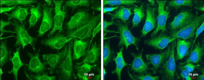
- Immunocytochemistry-Immunofluorescence analysis of DNase I was performed in HeLa cells fixed in ice cold MeOH for 5 min. Green: DNase I Polyclonal Antibody (Product # PA5 22017) diluted at 1:200. Blue: Hoechst 33342 staining. Scale bar = 10 µm.
- Submitted by
- Invitrogen Antibodies (provider)
- Main image

- Experimental details
- Immunocytochemistry-Immunofluorescence analysis of DNase I was performed in HeLa cells fixed in ice cold MeOH for 5 min. Green: DNase I Polyclonal Antibody (Product # PA5 22017) diluted at 1:200. Blue: Hoechst 33342 staining. Scale bar = 10 µm.

Supportive validation
- Submitted by
- Invitrogen Antibodies (provider)
- Main image
- Experimental details
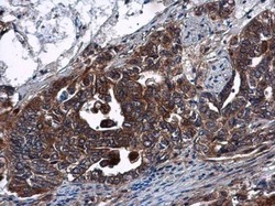
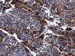
- DNase I Polyclonal Antibody detects DNase I protein at cytoplasm in human cervical carcinoma by immunohistochemical analysis. Sample: Paraffin-embedded human cervical carcinoma. DNase I Polyclonal Antibody (Product # PA5-22017) diluted at 1:500. Antigen Retrieval: Citrate buffer, pH 6.0, 15 min.
- Submitted by
- Invitrogen Antibodies (provider)
- Main image
- Experimental details
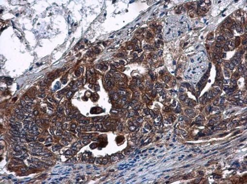
- DNase I Polyclonal Antibody detects DNase I protein at cytoplasm in human endometrial carcinoma by immunohistochemical analysis. Sample: Paraffin-embedded human endometrial carcinoma. DNase I Polyclonal Antibody (Product # PA5-22017) diluted at 1:500. Antigen Retrieval: Citrate buffer, pH 6.0, 15 min.

Supportive validation
- Submitted by
- Invitrogen Antibodies (provider)
- Main image
- Experimental details
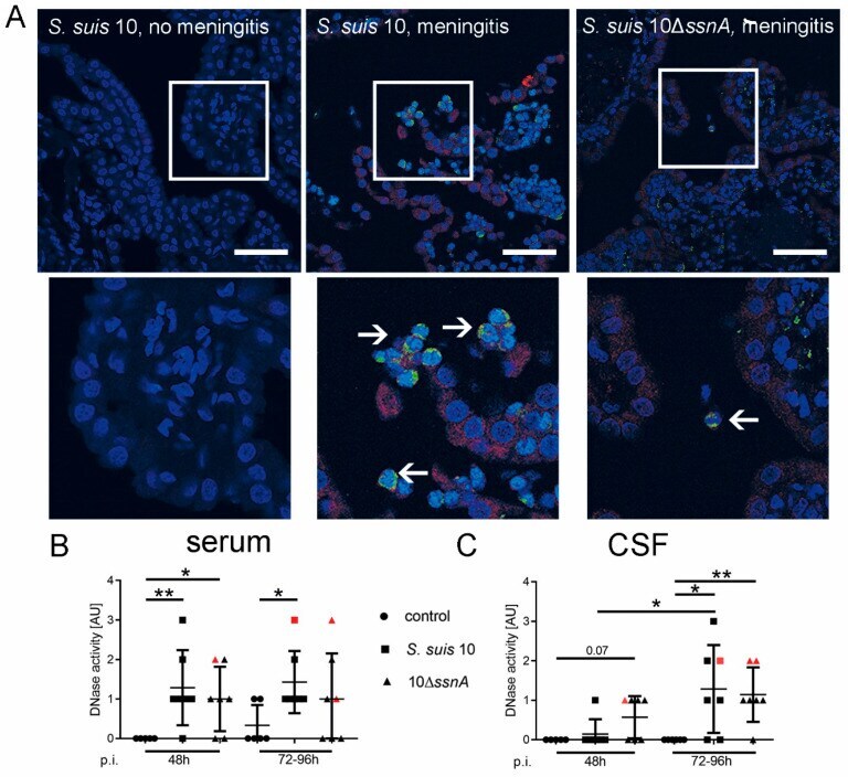
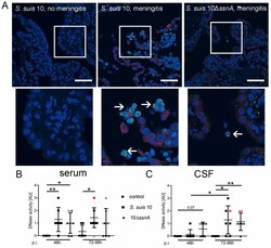
- Figure 4 Host DNases are detectable and active during S. suis meningitis in choroid plexus, CSF and serum. ( A ) By immunofluorescence staining, we could show that the amount of DNase 1 in the choroid plexus is different in animals with inflammation, compared to animals without inflammation of the choroid plexus. In the choroid plexus of an infected pig without clinical signs of CNS disorders only weak DNase 1 signals were detected in the early phase experiment. In pigs with clinical signs of CNS disorders a high DNase 1 signal in the region of the choroid plexus epithelial cells was observed. PR-39 signal, as a marker for neutrophils, was found in these pigs as well (blue = DNA (Hoechst); green = PR-39; red = DNase 1 (scale bar upper panel = 50 um; lower panel = 20 um; arrows mark transmigrating or already transmigrated PR-39 positive neutrophils). Activity of DNases was observed in serum ( B ) and in CSF ( C ) of all pigs of the early phase experiment. A significant increase of DNase activity post-infection was found in serum for both infection groups after 48 h and for the S. suis 10-infected group also after 96 h. In CSF after 96 h, a higher increase was observed than after 48 h in both infection groups. Data shown as mean +- SD. Statistical analysis: unpaired Mann-Whitney test (* p < 0.05, ** p < 0.01).