 Explore
 Validate
 Learn
LearnPA5-77396
antibody from Invitrogen Antibodies
Targeting: GPER1
CEPR, CMKRL2, DRY12, FEG-1, GPCR-Br, GPER, GPR30, LERGU, LERGU2, LyGPR
 Western blot
Antibody data
- Antibody Data
- Antigen structure
- References [1]
- Comments [0]
- Validations
- Western blot [3]
- Immunocytochemistry [1]
- Immunohistochemistry [1]
Submit
Validation data
Reference
Comment
Report error
- Product number
- PA5-77396 - Provider product page

- Provider
- Invitrogen Antibodies
- Product name
- GPR30 Polyclonal Antibody
- Antibody type
- Polyclonal
- Antigen
- Synthetic peptide
- Description
- For reconstitution, we recommend adding 100 µL distilled water to a final antibody concentration of about 1 mg/mL. To use this carrier-free antibody for conjugation experiments, we strongly recommend performing another round of desalting. (Zeba Spin Desalting Columns, 7KMWCO, 0.5 mL, Product # 89882)
- Reactivity
- Human, Mouse, Rat
- Host
- Rabbit
- Isotype
- IgG
- Vial size
- 50 µL
- Concentration
- 0.8 mg/mL
- Storage
- -20°C
Submitted references Essential and sex-specific effects of mGluR5 in ventromedial hypothalamus regulating estrogen signaling and glucose balance.
Fagan MP, Ameroso D, Meng A, Rock A, Maguire J, Rios M
Proceedings of the National Academy of Sciences of the United States of America 2020 Aug 11;117(32):19566-19577
Proceedings of the National Academy of Sciences of the United States of America 2020 Aug 11;117(32):19566-19577
No comments: Submit comment
Supportive validation
- Submitted by
- Invitrogen Antibodies (provider)
- Main image

- Experimental details
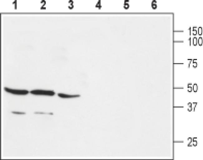
- Western blot analysis of rat brain (lanes 1 and 4), mouse brain (lanes 2 and 5) and rat lung (lanes 3 and 6) with GPR30 polyclonal antibody (Product # PA5-77396) using a dilution of 1:200.
- Submitted by
- Invitrogen Antibodies (provider)
- Main image

- Experimental details
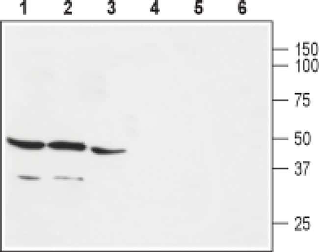
- Western blot analysis of rat brain (lanes 1 and 4), mouse brain (lanes 2 and 5) and rat lung (lanes 3 and 6) with GPR30 polyclonal antibody (Product # PA5-77396) using a dilution of 1:200.
- Submitted by
- Invitrogen Antibodies (provider)
- Main image
- Experimental details
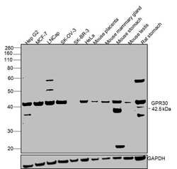
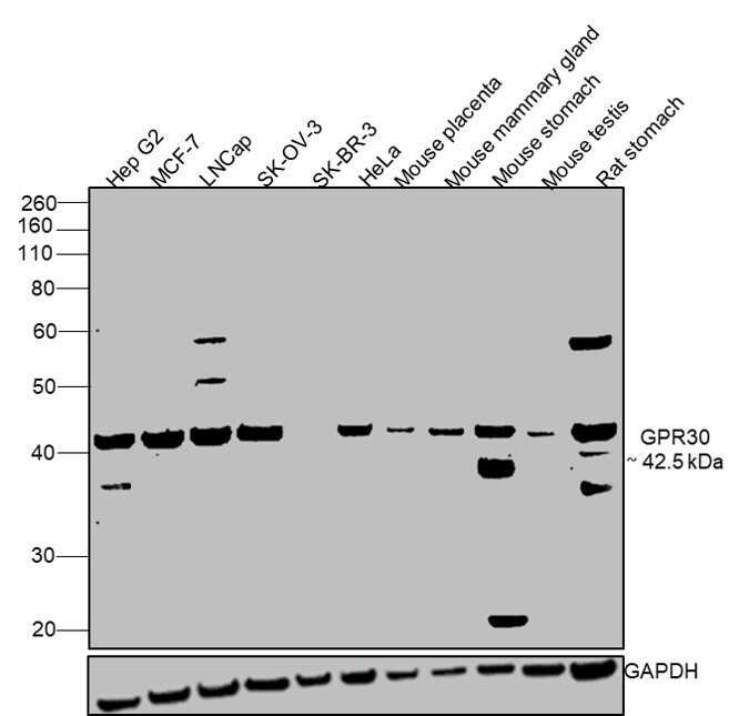
- Western blot was performed using anti-GPR30 Polyclonal Antibody (Product # PA5-77396) and a 42.5 kDa band corresponding to GPR30 was observed across all cell lines and tissues tested except SK-BR-3. Whole cell extracts (30 µg lysate) of Hep G2 (Lane 1), MCF-7 (Lane 2), LNCap (Lane 3), SK-OV-3 (Lane 4), SK-BR-3 (Lane 5), HeLa (Lane 6), Mouse placenta (Lane 7), Mouse mammary gland (Lane 8), Mouse stomach (Lane 9), Mouse testis (Lane 10), Rat stomach (Lane 11)were electrophoresed using NuPAGE® 4-12 % Bis-Tris gel (Product # NP0321BOX). Resolved proteins were then transferred onto a nitrocellulose membrane (Product # IB23001) by iBlot® 2 Dry Blotting System (Product # IB21001). The blot was probed with the primary antibody (1:2000 dilution) and detected by chemiluminescence with Goat anti-Rabbit IgG (H+L) Superclonal™ Recombinant Secondary Antibody, HRP (Product # A27036, 1:4000 dilution) using the iBright FL 1000 (Product # A32752). Chemiluminescent detection was performed using Novex® ECL Chemiluminescent Substrate Reagent Kit (Product # WP20005).
Supportive validation
- Submitted by
- Invitrogen Antibodies (provider)
- Main image

- Experimental details
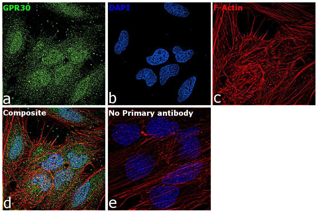
- Immunofluorescence analysis of GPR30 was performed using 70% confluent log phase HeLa cells. The cells were fixed with 4% paraformaldehyde for 10 minutes, permeabilized with 0.1% Triton™ X-100 for 15 minutes, and blocked with 2% BSA for 1 hour at room temperature. The cells were labeled with GPR30 Polyclonal Antibody (Product # PA5-77396) at 5µg/mL in 0.1% BSA, incubated at 4 degree Celsius overnight and then labeled with Goat anti-Rabbit IgG (H+L) Superclonal™ Recombinant Secondary Antibody, Alexa Fluor® 488 (Product # A27034) at a dilution of 1:2000 for 45 minutes at room temperature (Panel a: green). Nuclei (Panel b: blue) were stained with SlowFade® Gold Antifade Mountant with DAPI (Product # S36938). F-actin (Panel c: red) was stained with Rhodamine Phalloidin (Product # R415, 1:300). Panel d represents the merged image showing localization to cytoplasm and nucleus. Panel f represents control cells with no primary antibody to assess background. The images were captured at 60X magnification.
Supportive validation
- Submitted by
- Invitrogen Antibodies (provider)
- Main image
- Experimental details
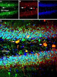
- Immunohistochemistry analysis of GPR30 in rat hippocampal dentate gyrus. A) Samples were probed with GPR30 polyclonal antibody (Product # PA5-77396) and incubated with parvalbumin (red) and DAPI. B) Nuclei stained image. C) Merged image of Panels A and B.
