Explore
 Validate
 Learn
LearnAF5534
antibody from R&D Systems
Targeting: GPER1
CEPR, CMKRL2, DRY12, FEG-1, GPCR-Br, GPER, GPR30, LERGU, LERGU2, LyGPR
 Western blot
Antibody data
- Antibody Data
- Antigen structure
- References [1]
- Comments [0]
- Validations
- Western blot [1]
- Immunohistochemistry [1]
- Flow cytometry [1]
Submit
Validation data
Reference
Comment
Report error
- Product number
- AF5534 - Provider product page

- Provider
- R&D Systems
- Product name
- Human GPER/GPR30 Antibody
- Antibody type
- Polyclonal
- Description
- Antigen Affinity-purified. Detects human GPER in direct ELISAs and Western blots. In direct ELISAs, less than 1% cross-reactivity with recombinant human GPR144, GPR115, and GPR124 is observed.
- Reactivity
- Human
- Host
- Goat
- Conjugate
- Unconjugated
- Antigen sequence
Q99527- Isotype
- IgG
- Vial size
- 100 ug
- Concentration
- LYOPH
- Storage
- Use a manual defrost freezer and avoid repeated freeze-thaw cycles. 12 months from date of receipt, -20 to -70 °C as supplied. 1 month, 2 to 8 °C under sterile conditions after reconstitution. 6 months, -20 to -70 °C under sterile conditions after reconstitution.
Submitted references G protein-coupled receptor 30 (GPR30) forms a plasma membrane complex with membrane-associated guanylate kinases (MAGUKs) and protein kinase A-anchoring protein 5 (AKAP5) that constitutively inhibits cAMP production.
Broselid S, Berg KA, Chavera TA, Kahn R, Clarke WP, Olde B, Leeb-Lundberg LM
The Journal of biological chemistry 2014 Aug 8;289(32):22117-27
The Journal of biological chemistry 2014 Aug 8;289(32):22117-27
No comments: Submit comment
Supportive validation
- Submitted by
- R&D Systems (provider)
- Main image
- Experimental details
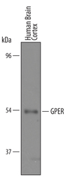
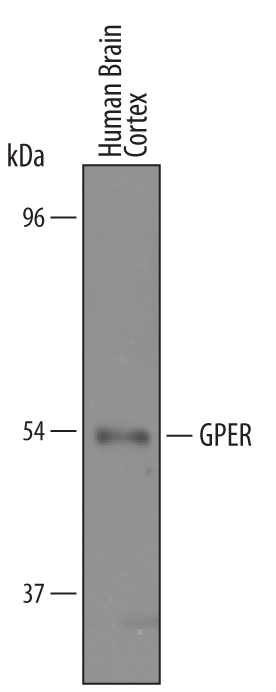
- Detection of Human GPER by Western Blot. Western blot shows lysates of human brain cortex tissue. PVDF membrane was probed with 1 µg/mL of Goat Anti-Human GPER Antigen Affinity-purified Polyclonal Antibody (Catalog # AF5534) followed by HRP-conjugated Anti-Goat IgG Secondary Antibody (Catalog # HAF019). A specific band was detected for GPER at approximately 54 kDa (as indicated). This experiment was conducted under reducing conditions and using Immunoblot Buffer Group 8.
Supportive validation
- Submitted by
- R&D Systems (provider)
- Main image
- Experimental details
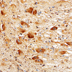
- GPER in Human Brain. GPER was detected in immersion fixed paraffin-embedded sections of human brain (hypothalamus) using Goat Anti-Human GPER Antigen Affinity-purified Polyclonal Antibody (Catalog # AF5534) at 10 µg/mL overnight at 4 °C. Tissue was stained using the Anti-Goat HRP-DAB Cell & Tissue Staining Kit (brown; Catalog # CTS008) and counterstained with hematoxylin (blue). Specific staining was localized to neuronal cell bodies. View our protocol for Chromogenic IHC Staining of Paraffin-embedded Tissue Sections.

Supportive validation
- Submitted by
- R&D Systems (provider)
- Main image
- Experimental details
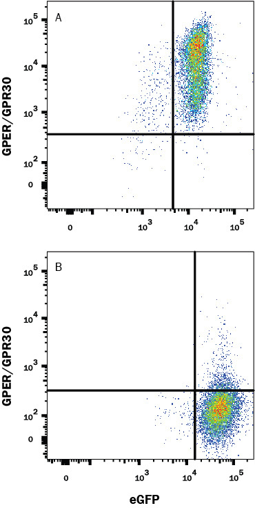
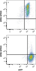
- Detection of GPER/GPR30 in HEK293 Human Cell Line Transfected with human GPER/GRP30 and eGFP by Flow Cytometry. HEK293 human embryonic kidney cell line transfected with (A) human GPER/GRP30 or (B) irrelevant transfectants, and eGFP was stained with Goat Anti-Human GPER/GPR30 Affinity-Purified Polyclonal Antibody (Catalog # AF5534) followed by Allophycocyanin-conjugated Anti-Goat IgG Secondary Antibody (Catalog # F0108). Quadrants were set based on Goat IgG Flow Cytometry Isotype Control (Catalog # AB-108-C, data not shown). View our protocol for Staining Membrane-associated Proteins.