Explore
 Validate
 Learn
Learn Western blot
Antibody data
- Antibody Data
- Antigen structure
- References [0]
- Comments [0]
- Validations
- Western blot [1]
- Immunocytochemistry [2]
- Immunohistochemistry [3]
Submit
Validation data
Reference
Comment
Report error
- Product number
- PA3-039 - Provider product page

- Provider
- Invitrogen Antibodies
- Product name
- GPR82 Polyclonal Antibody
- Antibody type
- Polyclonal
- Antigen
- Other
- Description
- IHC(P) analysis shows positive staining of GPR82 in human kidney tubules and germinal center of human lymph node. WB analysis shows GPR82 in glyco-protein enriched fractions from mock transfected or GPR82 overexpressed 293 cells. GPR82 exists in different glycosylated forms which may cause the molecular weight detected by WB to vary.
- Reactivity
- Human
- Host
- Rabbit
- Isotype
- IgG
- Vial size
- 100 μL
- Concentration
- Conc. Not Determined
- Storage
- -20°C, Avoid Freeze/Thaw Cycles
No comments: Submit comment
Supportive validation
- Submitted by
- Invitrogen Antibodies (provider)
- Main image

- Experimental details
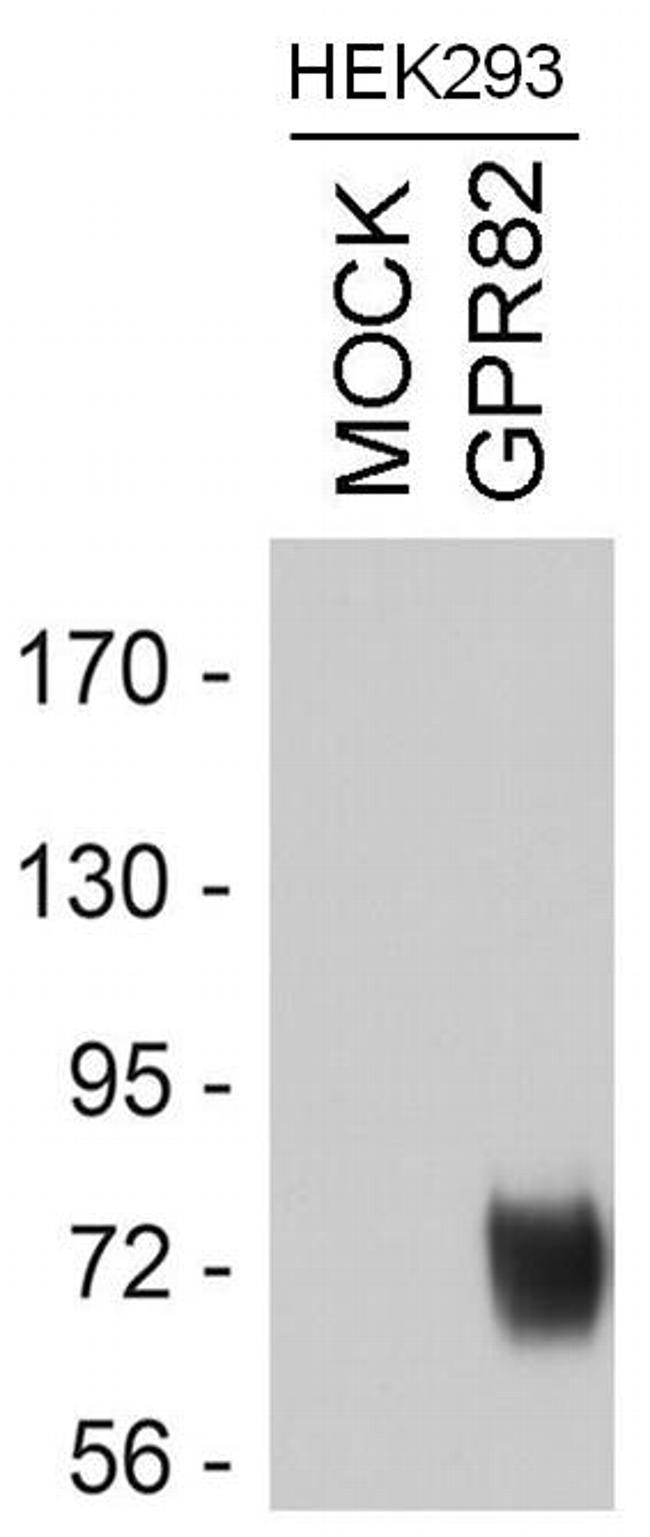
- Western blot analysis of GPR82 was performed by loading equal amounts of wheat germ lectin agarose bead enriched GPR receptor fractions from mock-transfected or GPR82 transfected HEK293 lysates onto a 7.5% Tris-HCl polyacrylamide gel. Proteins were transferred to a PVDF membrane, blocked and probed with a GPR82 polyclonal antibody (Product # PA3-039) at a dilution of 1:5000, overnight at 4C on a rocking platform, followed by an HRP-conjugated goat anti-rabbit IgG secondary antibody. Denatured GPR82 was detected at ~72kDa. Chemiluminescent detection was performed using ECL.
Supportive validation
- Submitted by
- Invitrogen Antibodies (provider)
- Main image

- Experimental details
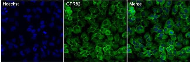
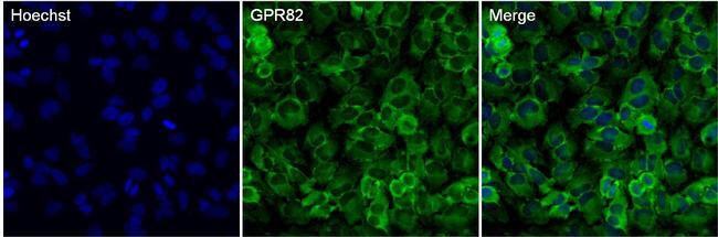
- Immunofluoresent analysis of GPR82 in HeLa cells. The cells were fixed with 4% paraformaldehyde in PBS for 15 minutes and blocked with 3% BSA in PBS for 30 minutes at room temperature. Cells were stained with a GPR82 rabbit polyclonal antibody (Product # PA3-039) at a dilution of 1:500 in blocking buffer for 1 hour at room temperature, and then incubated with Goat anti-Rabbit IgG (H+L) Secondary Antibody, Alexa Fluor® 488 conjugate (Product # A-11034) at a dilution of 1:1000 for at least 30 minutes at room temperature in the dark (green). Nuclei (blue) were stained with Hoechst 33342 (Product # 62249). Images were taken on a Thermo Scientific ToxInsight Instrument at 20X magnification.
- Submitted by
- Invitrogen Antibodies (provider)
- Main image
- Experimental details
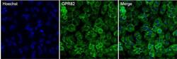
- Immunofluoresent analysis of GPR82 in HeLa cells. The cells were fixed with 4% paraformaldehyde in PBS for 15 minutes and blocked with 3% BSA in PBS for 30 minutes at room temperature. Cells were stained with a GPR82 rabbit polyclonal antibody (Product # PA3-039) at a dilution of 1:500 in blocking buffer for 1 hour at room temperature, and then incubated with Goat anti-Rabbit IgG (H+L) Secondary Antibody, Alexa Fluor® 488 conjugate (Product # A-11034) at a dilution of 1:1000 for at least 30 minutes at room temperature in the dark (green). Nuclei (blue) were stained with Hoechst 33342 (Product # 62249). Images were taken on a Thermo Scientific ToxInsight Instrument at 20X magnification.
Supportive validation
- Submitted by
- Invitrogen Antibodies (provider)
- Main image

- Experimental details
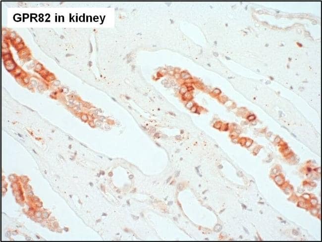
- Immunohistochemistry analysis of GPR82 was performed on human kidney tissue. To expose target proteins, antigen retrieval was performed by microwaving tissues for 20 minutes in 10mM sodium citrate buffer (pH 6.0). Tissue slides were probed with a GPR82 polyclonal antibody (Product # PA3-039) at a dilution of 1:3000, overnight at 4C in a humidified chamber. Tissues were washed, and detection was performed using an ABC kit composed of biotinylated goat anti-rabbit IgG, peroxidase-conjugated avidin, and 3-amino-9-ethylcarbazole (AEC) substrate in acetate buffer. Tissues were counterstained with hematoxylin and dehydrated to prep for mounting.

- Submitted by
- Invitrogen Antibodies (provider)
- Main image
- Experimental details
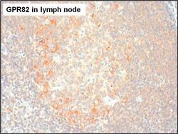
- Immunohistochemistry analysis of GPR82 was performed on human lymph node tissue. To expose target proteins, antigen retrieval was performed by microwaving tissues for 20 minutes in 10mM sodium citrate buffer (pH 6.0). Tissue slides were probed with a GPR82 polyclonal antibody (Product # PA3-039) at a dilution of 1:3000, overnight at 4C in a humidified chamber. Tissues were washed, and detection was performed using an ABC kit composed of biotinylated goat anti-rabbit IgG, peroxidase-conjugated avidin, and 3-amino-9-ethylcarbazole (AEC) substrate in acetate buffer. Tissues were counterstained with hematoxylin and dehydrated to prep for mounting.

- Submitted by
- Invitrogen Antibodies (provider)
- Main image

- Experimental details
- Immunohistochemistry analysis of GPR82 was performed on human kidney tissue. To expose target proteins, antigen retrieval was performed by microwaving tissues for 20 minutes in 10mM sodium citrate buffer (pH 6.0). Tissue slides were probed with a GPR82 polyclonal antibody (Product # PA3-039) at a dilution of 1:3000, overnight at 4C in a humidified chamber. Tissues were washed, and detection was performed using an ABC kit composed of biotinylated goat anti-rabbit IgG, peroxidase-conjugated avidin, and 3-amino-9-ethylcarbazole (AEC) substrate in acetate buffer. Tissues were counterstained with hematoxylin and dehydrated to prep for mounting.