Explore
 Validate
 Learn
Learn Western blot
 Immunoprecipitation
Antibody data
- Antibody Data
- Antigen structure
- References [1]
- Comments [0]
- Validations
- Western blot [2]
- Immunocytochemistry [2]
- Other assay [2]
Submit
Validation data
Reference
Comment
Report error
- Product number
- MA5-24769 - Provider product page

- Provider
- Invitrogen Antibodies
- Product name
- S100A10 Monoclonal Antibody (6F4-E6-D5-C10)
- Antibody type
- Monoclonal
- Antigen
- Purifed from natural sources
- Reactivity
- Human, Mouse, Rat
- Host
- Mouse
- Isotype
- IgG
- Antibody clone number
- 6F4-E6-D5-C10
- Vial size
- 100 µL
- Concentration
- 1 mg/mL
- Storage
- -20°C
Submitted references Neuron-Derived Estrogen Is Critical for Astrocyte Activation and Neuroprotection of the Ischemic Brain.
Lu Y, Sareddy GR, Wang J, Zhang Q, Tang FL, Pratap UP, Tekmal RR, Vadlamudi RK, Brann DW
The Journal of neuroscience : the official journal of the Society for Neuroscience 2020 Sep 16;40(38):7355-7374
The Journal of neuroscience : the official journal of the Society for Neuroscience 2020 Sep 16;40(38):7355-7374
No comments: Submit comment
Supportive validation
- Submitted by
- Invitrogen Antibodies (provider)
- Main image
- Experimental details
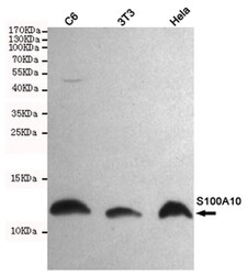
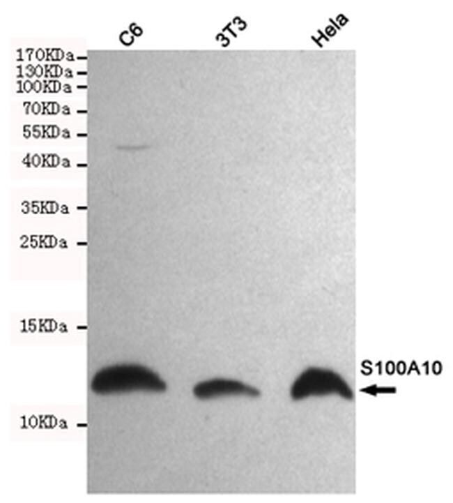
- Western blot analysis of S100a10 in C6, 3T3 and HeLa lysate. The membrane was probed with S100a10 monoclonal antibody (Product # MA5-24769) with a dilution of 1:1000.
- Submitted by
- Invitrogen Antibodies (provider)
- Main image
- Experimental details
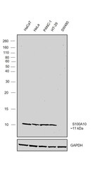
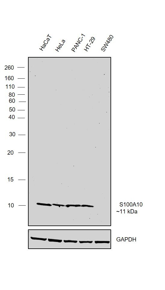
- Western Blot was performed using Anti-S100A10 Monoclonal Antibody (6F4-E6-D5-C10) (Product # MA5-24769) and a 11 kDa band corresponding to Protein S100-A10 was observed across the panel tested except SW480 which is reported to be negative. Whole cell extracts (30 µg lysate) of HaCaT (Lane 1), HeLa (Lane 2), PANC-1 (Lane 3), HT-29 (Lane 4), SW480 (Lane 5) were electrophoresed using NuPAGE™ 4-12% Bis-Tris Protein Gel (Product # NP0322BOX). Resolved proteins were then transferred onto a nitrocellulose membrane (Product # IB23001) by iBlot® 2 Dry Blotting System (Product # IB21001). The blot was probed with the primary antibody (1:1000 dilution) and detected by chemiluminescence with Goat anti-Mouse IgG (H+L) Superclonal™ Recombinant Secondary Antibody, HRP (Product # A28177, 1:4000 dilution) using the iBright FL 1000 (Product # A32752). Chemiluminescent detection was performed using Novex® ECL Chemiluminescent Substrate Reagent Kit (Product # WP20005).
Supportive validation
- Submitted by
- Invitrogen Antibodies (provider)
- Main image
- Experimental details
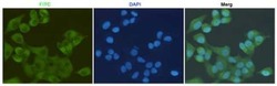
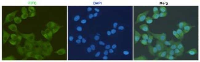
- Immunocytochemistry analysis of S100a10 in HeLa cells fixed with 4% paraformaldehyde. Cells were probed with a S100a10 monoclonal antibody (Product # MA5-24769) using a dilution of 1:200.
- Submitted by
- Invitrogen Antibodies (provider)
- Main image
- Experimental details
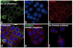
- Immunofluorescence analysis of Protein S100-A10 was performed using 70% confluent log phase HT-29 cells. The cells were fixed with 4% paraformaldehyde for 10 minutes, permeabilized with 0.1% Triton™ X-100 for 15 minutes, and blocked with 2% BSA for 45 minutes at room temperature. The cells were labeled with S100A10 Monoclonal Antibody (6F4-E6-D5-C10) (Product # MA5-24769) at 1:200 dilution in 0.1% BSA, incubated at 4 degree celsius overnight and then labeled with Donkey anti-Mouse IgG (H+L) Highly Cross-Adsorbed Secondary Antibody, Alexa Fluor Plus 488 (Product # A32766), (1:3000 dilution), for 45 minutes at room temperature (Panel a: Green). Nuclei (Panel b:Blue) were stained with ProLong™ Diamond Antifade Mountant with DAPI (Product # P36962). F-actin (Panel c: Red) was stained with Rhodamine Phalloidin (Product # R415, 1:300). Panel d represents the merged image showing cytoplasmic localization. Panel e represents merged image of SW480 cells showing no staining. Panel f represents control cells with no primary antibody to assess background. The images were captured at 60X magnification.

Supportive validation
- Submitted by
- Invitrogen Antibodies (provider)
- Main image
- Experimental details
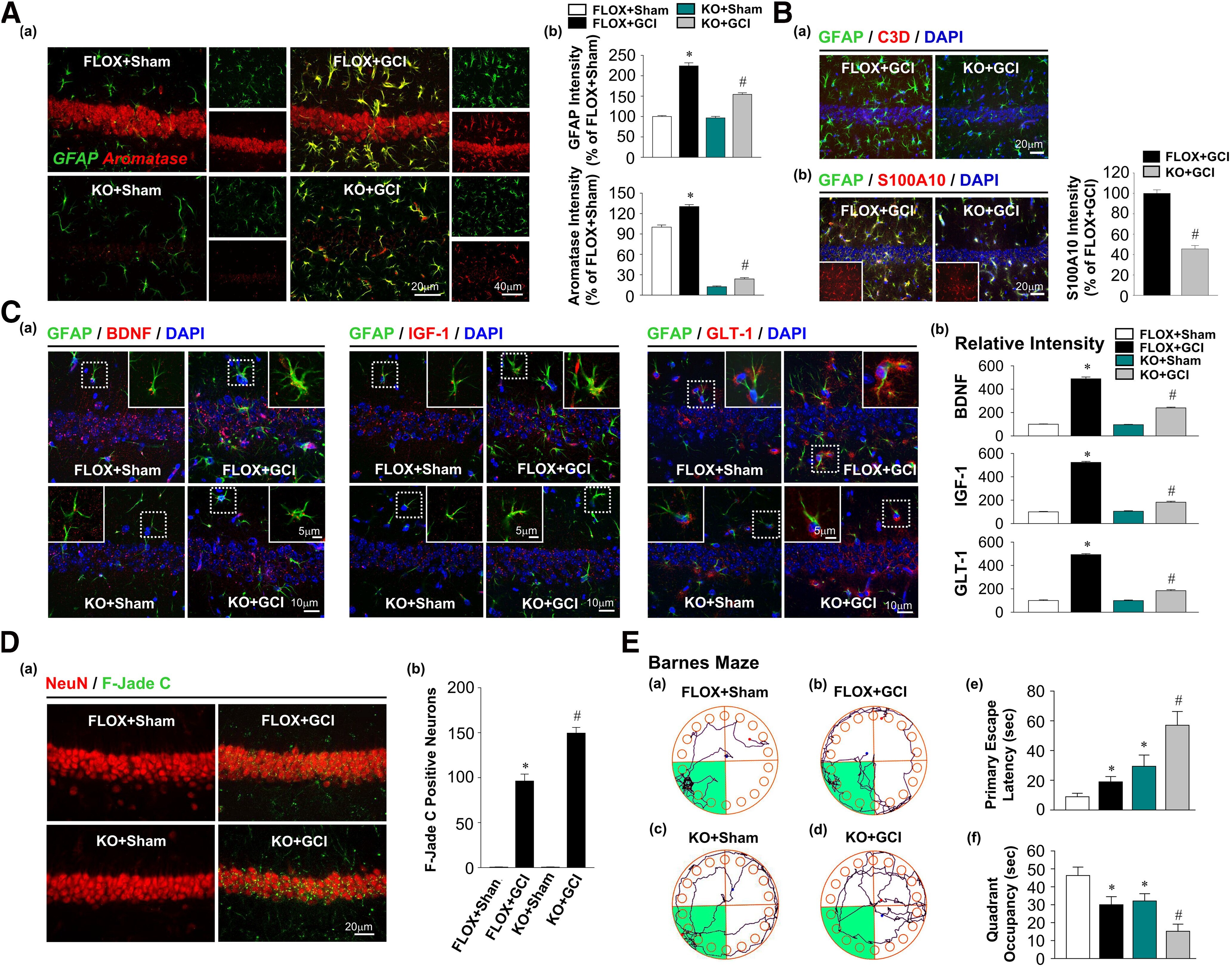
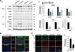
- Figure 4. Ovariectomized female FBN-ARO-KO mice have diminished astrocyte A2 phenotype 7 d after GCI. Aa , Astrocyte PAN-reactive, A1-specific, and A2-specific phenotypes were determined by Western blot analysis with purified astrocyte lysates from ovariectomized female brains 7 d after GCI reperfusion. Ab , Levels of the examined markers for different astrocyte phenotypes were quantitatively analyzed. N = 3. B , IHC analysis for astrocyte A1 phenotype by double staining of the selected astrocyte A1 marker C3D with GFAP. Ca , IHC examination for astrocyte A2 phenotype using A2-specific marker S100A10 and GFAP double staining. Cb , Percentage changes of S100A10 in FBN-ARO-KO+GCI mice versus FLOX+GCI mice were quantified. N = 4. Values are mean +- SEM of determinations from each group. * p < 0.05 versus FLOX+Sham. # p < 0.05 versus FLOX+GCI. ND, Nondetectable.
- Submitted by
- Invitrogen Antibodies (provider)
- Main image
- Experimental details
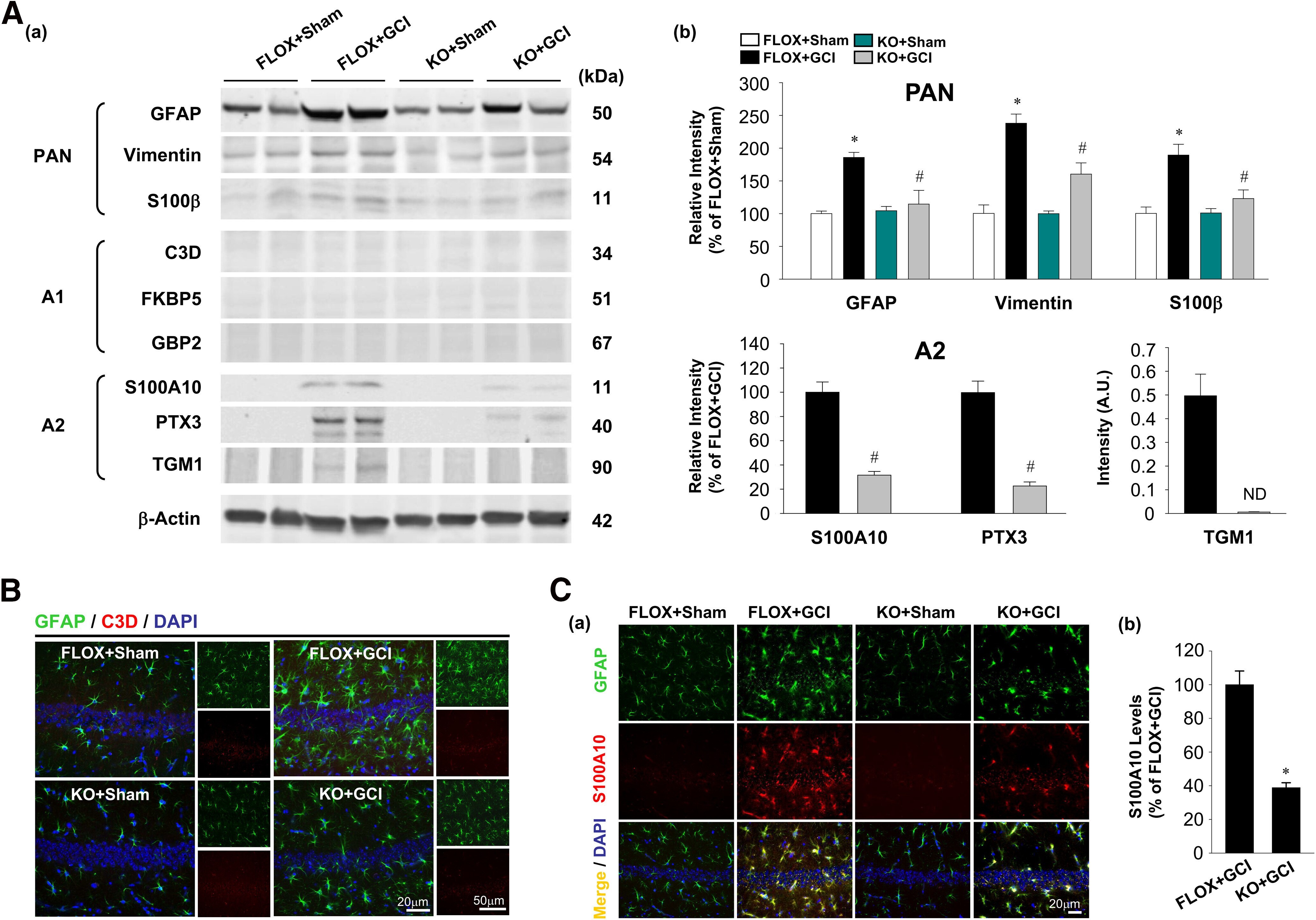
- Figure 7. Male FBN-ARO-KO mice exhibit strong neurodegeneration and cognitive impairment 7 d after GCI injury. Aa , Astrocyte activation and aromatization in hippocampal CA1 region were examined by GFAP and aromatase double staining. Ab , GFAP levels in each group were quantified as a parameter of astrocyte reactivity after GCI injury. Total aromatase levels in hippocampal CA1 region were further measured. B , Astrocyte A1 and A2 phenotypes after GCI were examined by IHC analysis with the markers of C3D ( Ba ) and S100A10 ( Bb ), respectively. Ca , BDNF, IGF-1, and GLT-1 production in astrocytes following the altered astrocyte activation was determined by IHC analysis. Cb , Their relative changes of intensities in astrocytes were quantified. Da , Representative double staining for NeuN and F-Jade C to evaluate neuronal degeneration in each group. Db , F-Jade C-positive neurons were counted. E , Spatial reference memory was assessed by Barnes Maze behavioral test. Ea-Ed , Tracking plots in probe trial. Primary escape latency ( Ee ) and quadrant occupancy ( Ef ) of animals from each group in probe trial were recorded. Values are mean +- SEM of determinations from each group. N = 4 for A-C ; N = 8-11 for D . * p < 0.05 versus FLOX+Sham. # p < 0.05 versus FLOX+GCI.