Explore
 Validate
 Learn
Learn Western blot
 ELISA
ELISA Immunocytochemistry
Antibody data
- Antibody Data
- Antigen structure
- References [1]
- Comments [0]
- Validations
- Immunocytochemistry [1]
- Other assay [1]
Submit
Validation data
Reference
Comment
Report error
- Product number
- PA1-28875 - Provider product page

- Provider
- Invitrogen Antibodies
- Product name
- TFF1 Polyclonal Antibody
- Antibody type
- Polyclonal
- Antigen
- Synthetic peptide
- Description
- Recommended positive controls: About 70% of breast carcinomas show partial staining of tumor cells..
- Reactivity
- Human
- Host
- Rabbit
- Isotype
- IgG
- Vial size
- 250 μL
- Concentration
- Conc. Not Determined
- Storage
- 4°C, do not freeze
Submitted references Gastric Proteins MUC5AC and TFF1 as Potential Diagnostic Markers of Colonic Sessile Serrated Adenomas/Polyps.
Khaidakov M, Lai KK, Roudachevski D, Sargsyan J, Goyne HE, Pai RK, Lamps LW, Hagedorn CH
American journal of clinical pathology 2016 Nov 1;146(5):530-537
American journal of clinical pathology 2016 Nov 1;146(5):530-537
No comments: Submit comment
Supportive validation
- Submitted by
- Invitrogen Antibodies (provider)
- Main image

- Experimental details
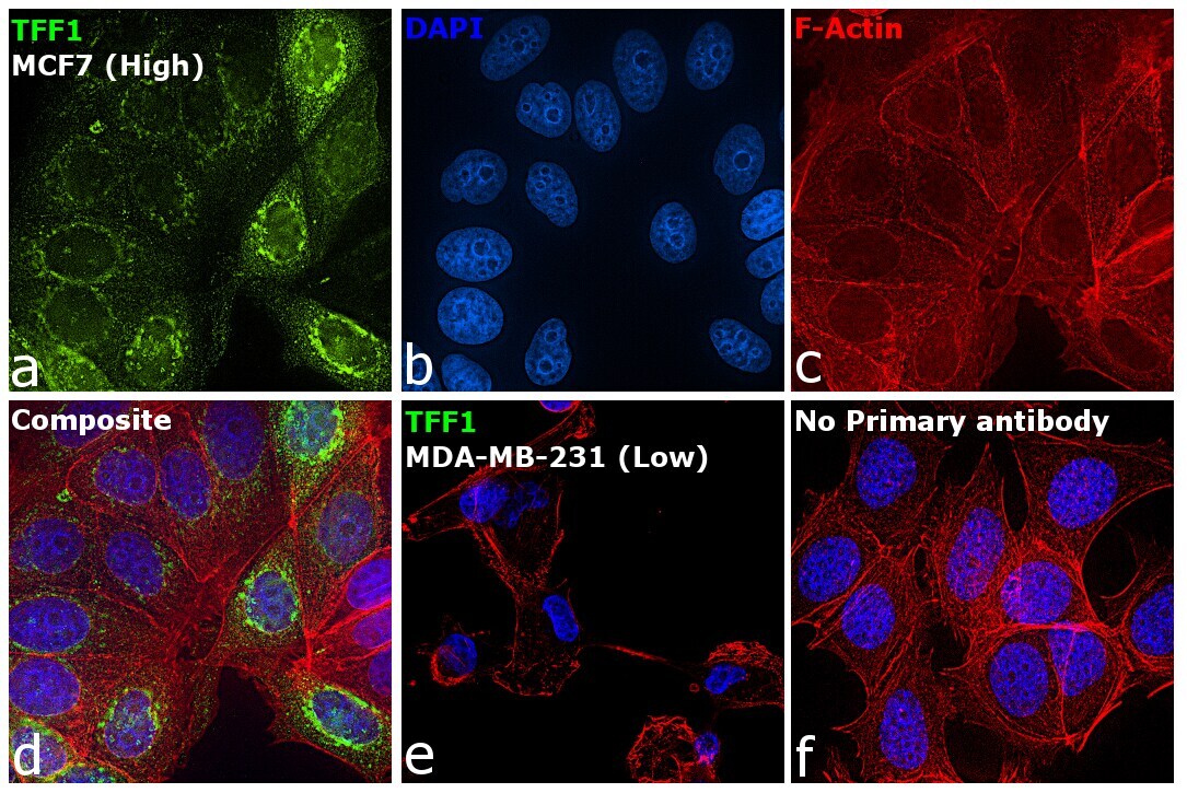
- Immunofluorescence analysis of Trefoil factor 1 was performed using 70% confluent log phase MCF7 cells. The cells were fixed with 4% paraformaldehyde for 10 minutes, permeabilized with 0.1% Triton™ X-100 for 15 minutes, and blocked with 2% BSA for 45 minutes at room temperature. The cells were labeled with TFF1 Polyclonal Antibody (Product # PA1-28875) at 1:100 dilution in 0.1% BSA, incubated at 4 degree celsius overnight and then labeled with Donkey anti-Rabbit IgG (H+L) Highly Cross-Adsorbed Secondary Antibody, Alexa Fluor Plus 488 (Product # A32790), (1:2000 dilution), for 45 minutes at room temperature (Panel a: Green). Nuclei (Panel b: Blue) were stained with ProLong™ Diamond Antifade Mountant with DAPI (Product # P36962). F-actin (Panel c: Red) was stained with Rhodamine Phalloidin (Product # R415, 1:300). Panel d represents the merged image showing cytoplasmic localization. Panel e represents MDA-MB-231 cells showing no expression of TFF1. Panel f represents control cells with no primary antibody to assess background. The images were captured at 60X magnification.
Supportive validation
- Submitted by
- Invitrogen Antibodies (provider)
- Main image
- Experimental details
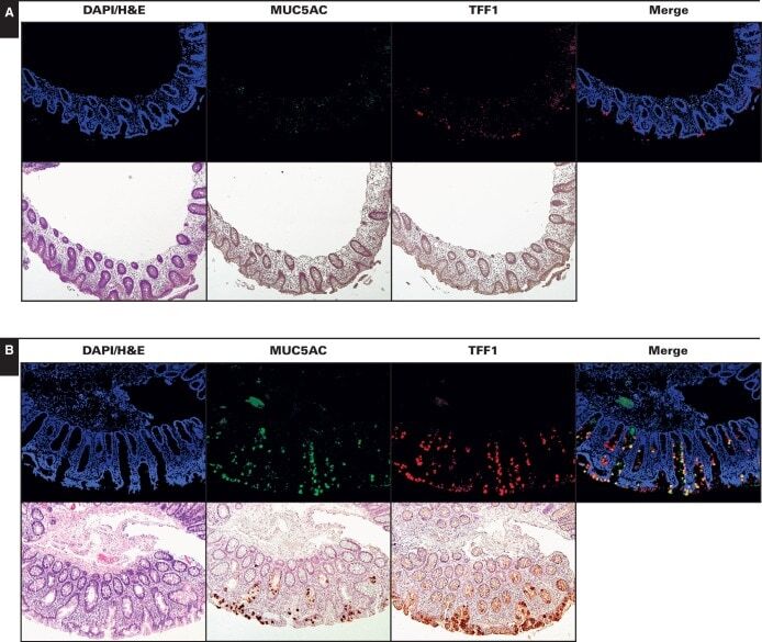
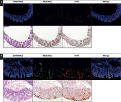
- Image 1 Expression of MUC5AC and TFF1 in normal colon ( A ), hyperplastic polyps (HPs) ( B ), and sessile serrated adenomas/polyps (SSA/Ps) ( C ) (x10). Immunofluorescence and immunoperoxidase staining of the same region is shown for representative control (normal), HP, and SSA/P samples. As expected, the signals for gastric proteins MUC5AC and TFF1 were negligible in normal colonic mucosa. MUC5AC and TFF1 immunopositive cells were present in HPs. In SSA/Ps, MUC5AC and TFF1 were typically expressed by most of the epithelial cells along the entire length of architecturally compromised crypts. SSA/Ps, compared with HPs, also showed significant coexpression of both MUC5AC and TFF1 in merged immunofluorescence analyses.