 Explore
 Validate
 Learn
Learn Western blot
 Immunocytochemistry
Antibody data
- Antibody Data
- Antigen structure
- References [2]
- Comments [0]
- Validations
- Immunocytochemistry [1]
- Immunohistochemistry [2]
- Other assay [2]
Submit
Validation data
Reference
Comment
Report error
- Product number
- PA5-31863 - Provider product page

- Provider
- Invitrogen Antibodies
- Product name
- TFF1 Polyclonal Antibody
- Antibody type
- Polyclonal
- Antigen
- Recombinant full-length protein
- Description
- Recommended positive controls: MCF-7. Store product as a concentrated solution. Centrifuge briefly prior to opening the vial.
- Reactivity
- Human, Mouse
- Host
- Rabbit
- Isotype
- IgG
- Vial size
- 100 μL
- Concentration
- 0.17 mg/mL
- Storage
- Store at 4°C short term. For long term storage, store at -20°C, avoiding freeze/thaw cycles.
Submitted references TFF-1 Functions to Suppress Multiple Phenotypes Associated with Lung Cancer Progression.
Heparanase Promotes Syndecan-1 Expression to Mediate Fibrillar Collagen and Mammographic Density in Human Breast Tissue Cultured ex vivo.
Minegishi K, Dobashi Y, Tsubochi H, Hagiwara K, Ishibashi Y, Nomura S, Nakamura R, Ohmoto Y, Endo S
OncoTargets and therapy 2021;14:4761-4777
OncoTargets and therapy 2021;14:4761-4777
Heparanase Promotes Syndecan-1 Expression to Mediate Fibrillar Collagen and Mammographic Density in Human Breast Tissue Cultured ex vivo.
Huang X, Reye G, Momot KI, Blick T, Lloyd T, Tilley WD, Hickey TE, Snell CE, Okolicsanyi RK, Haupt LM, Ferro V, Thompson EW, Hugo HJ
Frontiers in cell and developmental biology 2020;8:599
Frontiers in cell and developmental biology 2020;8:599
No comments: Submit comment
Supportive validation
- Submitted by
- Invitrogen Antibodies (provider)
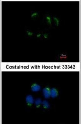
- Main image
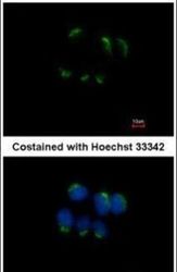
- Experimental details
- Immunofluorescent analysis of Trefoil factor 1 in methanol-fixed MCF-7 cells using a Trefoil factor 1 polyclonal antibody (Product # PA5-31863) at a 1:500 dilution.
Supportive validation
- Submitted by
- Invitrogen Antibodies (provider)
- Main image
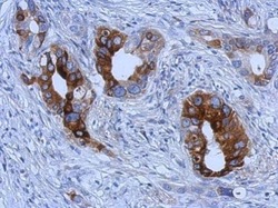
- Experimental details
- Immunohistochemical analysis of paraffin-embedded human gastric cancer, using Trefoil factor 1 (Product # PA5-31863) antibody at 1:500 dilution. Antigen Retrieval: EDTA based buffer, pH 8.0, 15 min.

- Submitted by
- Invitrogen Antibodies (provider)
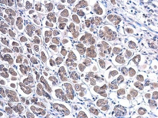
- Main image

- Experimental details
- Immunohistochemistry (Paraffin) analysis of TFF1 was performed in paraffin-embedded mouse stomach tissue using TFF1 Polyclonal Antibody (Product # PA5-31863) at a dilution of 1:500.
Supportive validation
- Submitted by
- Invitrogen Antibodies (provider)
- Main image
- Experimental details
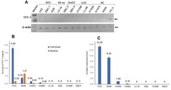
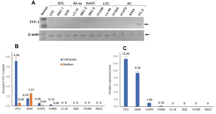
- Figure 1 Expression of endogenous TFF-1 protein in cultured cells from human lung carcinoma. ( A ) Expression of the TFF-1 protein was detected at around 13kDa by immunoblotting analysis only in PC-3 cells. ( B ) Schematic presentation of TFF-1 expression and the amount secreted into the medium analyzed by ELISA. ( C ) Result of qRT-PCR analysis of TFF-1 mRNA expression, expressed relative to values obtained for H1975. ( B and C ) Square and bar indicate mean value and standard deviation, respectively, obtained from three replicated experiments with three replicate samples. Abbreviations : AC, adenocarcinoma; SCC, squamous cell carcinoma; LCC, large cell carcinoma; SmCC, small cell carcinoma; Marker, molecular weight marker.
- Submitted by
- Invitrogen Antibodies (provider)
- Main image
- Experimental details
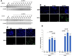
- Figure 2 Establishment of TFF-1-overexpressing cell lines. SBC-5 and LC-AI cells were transfected with a vector containing the cDNA encoding TFF-1/FLAG (pBA-TFF1) or empty vector (pBA-neo). ( A ) Expression of this fusion protein was evaluated by immunoblotting analysis with anti-TFF-1 antibody. Expression was not observed in parental SBC-5 or LC-AI cells (BC), but a single band of around 15kDa was detected after transfection of pBA-TFF1 (G-TFF1) in 4 clones, but not observed in cells transfected with pBA-neo (G-neo). Expression was normalized to the beta-actin loading control. ""Marker"" indicates molecular weight marker. ( B and C ) Images of immunofluorescent staining in parental cells (BC), pBA-neo (G-neo) or pBA-TFF1-transfected cells (G-TFF1) of SBC-5 ( B ), LC-AI ( C ) cells are shown. Cells were stained with anti-TFF-1 antibody and with 4',6-diamidino-2-phenylindole (DAPI). Scale bar indicates 100mum. ( D ) Schematic presentation of TFF-1 mRNA expression after transfection with pBA-neo or pBA-TFF1 (3 clones; CL) expressed relative to values obtained for H1975. Square and bar indicate mean values and standard deviations, respectively, obtained by qRT-PCR analysis from three replicated experiments with three replicate samples. Results were statistically analyzed and p-values were obtained by Fisher's PLSD test.
