Explore
 Validate
 Learn
Learn Flow cytometry
Antibody data
- Antibody Data
- Antigen structure
- References [5]
- Comments [0]
- Validations
- Flow cytometry [1]
- Other assay [1]
Submit
Validation data
Reference
Comment
Report error
- Product number
- 46-9490-42 - Provider product page

- Provider
- Invitrogen Antibodies
- Product name
- CD49a (Integrin alpha 1) Monoclonal Antibody (TS2/7), PerCP-eFluor™ 710, eBioscience™
- Antibody type
- Monoclonal
- Antigen
- Other
- Description
- Description: The monoclonal antibody TS2/7 reacts with human CD49a, also known as integrin alpha 1. CD49a is always associated with CD29 (integrin beta 1) to form a type I transmembrane protein called very late antigen-1 (VLA1). This heterodimer is expressed by endothelial cells, activated T cells, monocytes, a subset of NK cells, a subset of memory T cells, smooth muscle cells, and mesenchymal stem cells. CD49a binds to collagen IV strongly, but also binds to collagen I, laminin, and semaphorin7A. CD49a is involved in cell survival, proliferation, homing, and inflammatory responses. Applications Reported: This TS2/7 antibody has been reported for use in flow cytometric analysis. Applications Tested: This TS2/7 antibody has been pre-titrated and tested by flow cytometric analysis of stimulated normal human peripheral blood cells. This can be used at 5 µL (0.125 µg) per test. A test is defined as the amount (µg) of antibody that will stain a cell sample in a final volume of 100 µL. Cell number should be determined empirically but can range from 10^5 to 10^8 cells/test. PerCP-eFluor® 710 emits at 710 nm and is excited with the blue laser (488 nm); it can be used in place of PerCP-Cyanine5.5. We recommend using a 710/50 bandpass filter, however, the 695/40 bandpass filter is an acceptable alternative. Please make sure that your instrument is capable of detecting this fluorochrome. Light sensitivity: This tandem dye is sensitive to photo-induced oxidation. Please protect this vial and stained samples from light. Fixation: Samples can be stored in IC Fixation Buffer (Product # 00-8222) (100 µL of cell sample + 100 µL of IC Fixation Buffer) or 1-step Fix/Lyse Solution (Product # 00-5333) for up to 3 days in the dark at 4°C with minimal impact on brightness and FRET efficiency/compensation. Some generalizations regarding fluorophore performance after fixation can be made, but clone specific performance should be determined empirically. Excitation: 488 nm; Emission: 710 nm; Laser: Blue Laser. Filtration: 0.2 µm post-manufacturing filtered.
- Reactivity
- Human
- Host
- Mouse
- Isotype
- IgG
- Antibody clone number
- TS2/7
- Vial size
- 100 Tests
- Concentration
- 5 µL/Test
- Storage
- 4° C, store in dark, DO NOT FREEZE!
Submitted references Cancer stem-like cells evade CD8(+)CD103(+) tumor-resident memory T (T(RM)) lymphocytes by initiating an epithelial-to-mesenchymal transition program in a human lung tumor model.
α1β1 integrin-mediated adhesion inhibits macrophage exit from a peripheral inflammatory lesion.
alpha1beta1 Integrin+ and regulatory Foxp3+ T cells constitute two functionally distinct human CD4+ T cell subsets oppositely modulated by TNFalpha blockade.
Expression of the alpha1beta1 integrin, VLA-1, marks a distinct subset of human CD4+ memory T cells.
Glycoproteins of 210,000 and 130,000 m.w. on activated T cells: cell distribution and antigenic relation to components on resting cells and T cell lines.
Corgnac S, Damei I, Gros G, Caidi A, Terry S, Chouaib S, Deloger M, Mami-Chouaib F
Journal for immunotherapy of cancer 2022 Apr;10(4)
Journal for immunotherapy of cancer 2022 Apr;10(4)
α1β1 integrin-mediated adhesion inhibits macrophage exit from a peripheral inflammatory lesion.
Becker HM, Rullo J, Chen M, Ghazarian M, Bak S, Xiao H, Hay JB, Cybulsky MI
Journal of immunology (Baltimore, Md. : 1950) 2013 Apr 15;190(8):4305-14
Journal of immunology (Baltimore, Md. : 1950) 2013 Apr 15;190(8):4305-14
alpha1beta1 Integrin+ and regulatory Foxp3+ T cells constitute two functionally distinct human CD4+ T cell subsets oppositely modulated by TNFalpha blockade.
Goldstein I, Ben-Horin S, Koltakov A, Chermoshnuk H, Polevoy V, Berkun Y, Amariglio N, Bank I
Journal of immunology (Baltimore, Md. : 1950) 2007 Jan 1;178(1):201-10
Journal of immunology (Baltimore, Md. : 1950) 2007 Jan 1;178(1):201-10
Expression of the alpha1beta1 integrin, VLA-1, marks a distinct subset of human CD4+ memory T cells.
Goldstein I, Ben-Horin S, Li J, Bank I, Jiang H, Chess L
The Journal of clinical investigation 2003 Nov;112(9):1444-54
The Journal of clinical investigation 2003 Nov;112(9):1444-54
Glycoproteins of 210,000 and 130,000 m.w. on activated T cells: cell distribution and antigenic relation to components on resting cells and T cell lines.
Hemler ME, Sanchez-Madrid F, Flotte TJ, Krensky AM, Burakoff SJ, Bhan AK, Springer TA, Strominger JL
Journal of immunology (Baltimore, Md. : 1950) 1984 Jun;132(6):3011-8
Journal of immunology (Baltimore, Md. : 1950) 1984 Jun;132(6):3011-8
No comments: Submit comment
Supportive validation
- Submitted by
- Invitrogen Antibodies (provider)
- Main image
- Experimental details
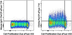
- Normal human peripheral blood cells were labeled with Cell Proliferation Dye eFluor® 450 (Product # 65-0842-85) then stimulated with Anti-Human CD3 Functional Grade Purified (Product # 16-0037-81), Anti-Human CD28 Functional Grade Purified (Product # 16-0289-81), and Human IL-2 Recombinant Protein (Product # 14-8029-81) for 7 days. Cells were then stained with Mouse IgG1 K Isotype Control PerCP-eFluor® 710 (Product # 46-4714-82) (left) or Anti-Human CD49a (Integrin alpha 1) PerCP-eFluor® 710 (right). Viable cells, as determined by Fixable Viability Dye eFluor® 506 (Product # 65-0866-14), were used for analysis.

Supportive validation
- Submitted by
- Invitrogen Antibodies (provider)
- Main image
- Experimental details
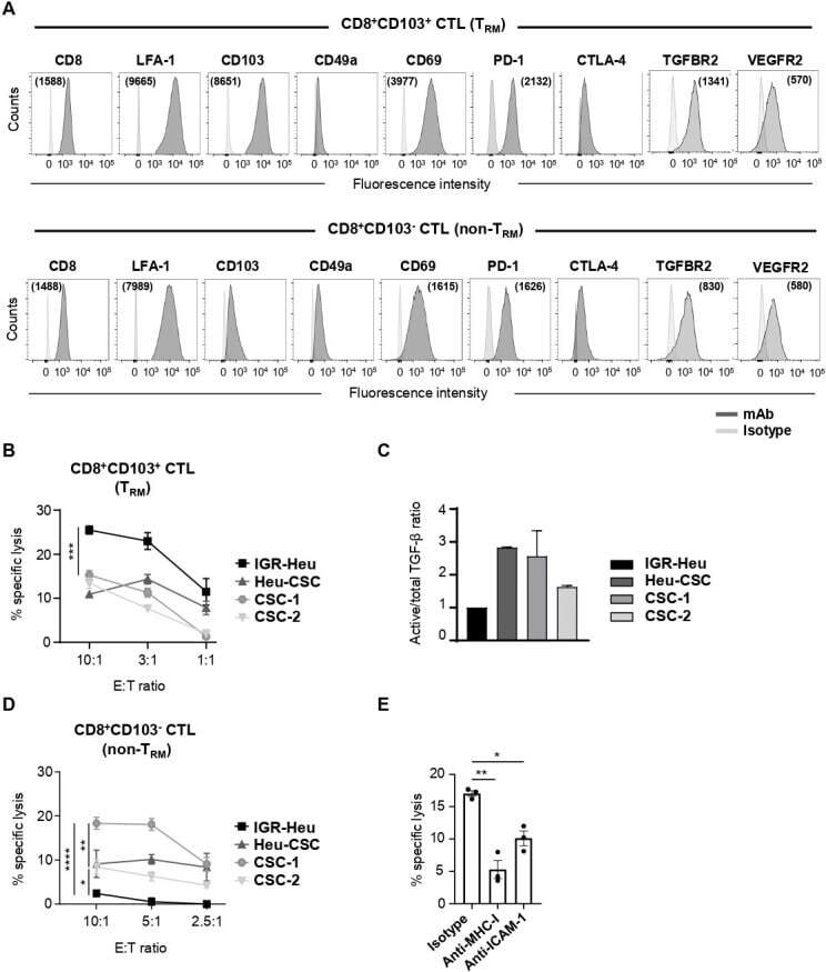
- Resident memory T (T RM ) and non-T RM phenotypic profiles and susceptibility of cancer stem cell (CSC) to cytotoxic T lymphocyte (CTL)-mediated killing. (A) Flow cytometry analyses of CD8, LFA-1, CD103, CD49a, CD69, PD-1, CTLA-4, TGFBR2 and VEGFR2 on CD8 + CD103 + T RM (Heu171) and CD8 + CD103 - non-T RM (H32-22) clones. Mean immunofluorescence intensity (MFI) are in parentheses. (B) Cytotoxic activity of the T RM clone (Heu171) towards autologous IGR-Heu, Heu-CSC, CSC-1 and CSC-2 target cells. Percent of specific lysis are shown at indicated effector to target (E:T) ratios. (C) Quantification of transforming growth factor (TGF)-beta in conditioned media from IGR-Heu, Heu-CSC, CSC-1 and CSC-2 by multi-analyte flow assay. Ratios of active/total TGF-beta normalized to IGR-Heu are included. Results are presented as mean+-SEM of duplicates. (D) Cytotoxicity of the non-T RM clone (H32-22) towards autologous IGR-Heu, Heu-CSC, CSC-1 and CSC-2 target cells. (E) Inhibition of T RM -cell-mediated killing. CSC-1 cells are preincubated in the presence of isotype control, anti-major histocompatibility complex class I (MHC-I) or anti-intercellular adhesion molecule 1 (ICAM-1) neutralizing monoclonal antibody (mAb) and then CTL were added at 5:1 E:T ratio. Symbols represent replicates and horizontal bars represent means+-SEM (n=3). P value was determined by two-way analysis of variance test. *p