Explore
 Validate
 Learn
LearnPA5-51662
antibody from Invitrogen Antibodies
Targeting: RAP1GAP
KIAA0474, RAP1GA1, RAP1GAP1, RAP1GAPII
 Immunocytochemistry
Antibody data
- Antibody Data
- Antigen structure
- References [0]
- Comments [0]
- Validations
- Immunocytochemistry [1]
- Immunohistochemistry [8]
Submit
Validation data
Reference
Comment
Report error
- Product number
- PA5-51662 - Provider product page

- Provider
- Invitrogen Antibodies
- Product name
- RAP1GAP Polyclonal Antibody
- Antibody type
- Polyclonal
- Antigen
- Recombinant full-length protein
- Description
- Immunogen sequence: IGIENIQEVQ EKRESPPAGQ KTPDSGHVSQ EPKSENSSTQ SSPEMPTTKN RAETAAQRAE ALKDFSRSSS SASSFASVVE ETEGVDGEDT GLESVSSSGT P Highest antigen sequence identity to the following orthologs: Mouse - 92%, Rat - 91%.
- Reactivity
- Human, Mouse
- Host
- Rabbit
- Isotype
- IgG
- Vial size
- 100 µL
- Concentration
- 0.3 mg/mL
- Storage
- Store at 4°C short term. For long term storage, store at -20°C, avoiding freeze/thaw cycles.
No comments: Submit comment
Supportive validation
- Submitted by
- Invitrogen Antibodies (provider)
- Main image
- Experimental details
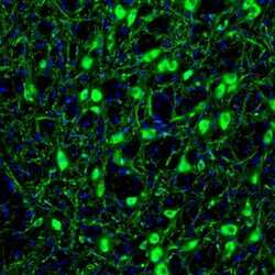
- Immunofluorescent staining of RAP1GAP in mouse globus pallidus shows strong cytoplasmic immunoreactivity in neurons. Samples were probed using a RAP1GAP Polyclonal Antibody (Product # PA5-51662).

Supportive validation
- Submitted by
- Invitrogen Antibodies (provider)
- Main image

- Experimental details
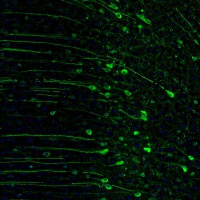
- Immunofluorescent staining of RAP1GAP in mouse midbrain shows strong immunoreactivity in locus coeruleus neurons. Samples were probed using a RAP1GAP Polyclonal Antibody (Product # PA5-51662).

- Submitted by
- Invitrogen Antibodies (provider)
- Main image

- Experimental details
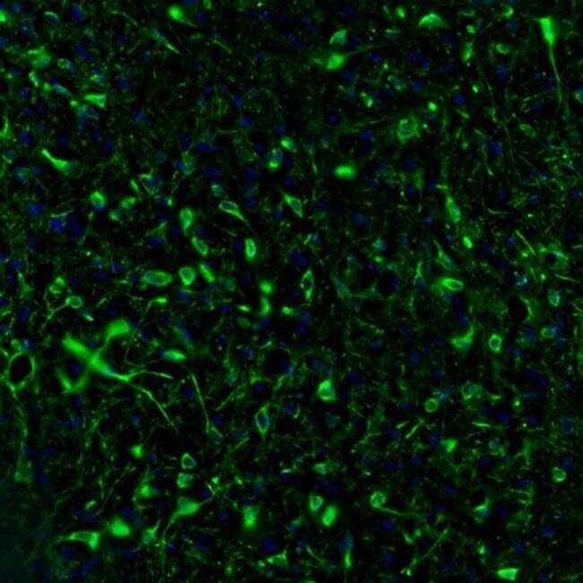
- Immunofluorescent staining of RAP1GAP in mouse cingulate cortex shows strong cytoplasmic positivity in neuronal cell bodies and proximal dendrites. Samples were probed using a RAP1GAP Polyclonal Antibody (Product # PA5-51662).
- Submitted by
- Invitrogen Antibodies (provider)
- Main image

- Experimental details
- Immunofluorescent staining of RAP1GAP in mouse hypothalamus shows positivity in neuronal cell bodies and processes in the lateral preoptic area. Samples were probed using a RAP1GAP Polyclonal Antibody (Product # PA5-51662).
- Submitted by
- Invitrogen Antibodies (provider)
- Main image

- Experimental details
- Immunofluorescent staining of RAP1GAP in mouse hippocampus shows cytoplasmic positivity in a subset of interneurons in the CA1 area. Samples were probed using a RAP1GAP Polyclonal Antibody (Product # PA5-51662).

- Submitted by
- Invitrogen Antibodies (provider)
- Main image

- Experimental details
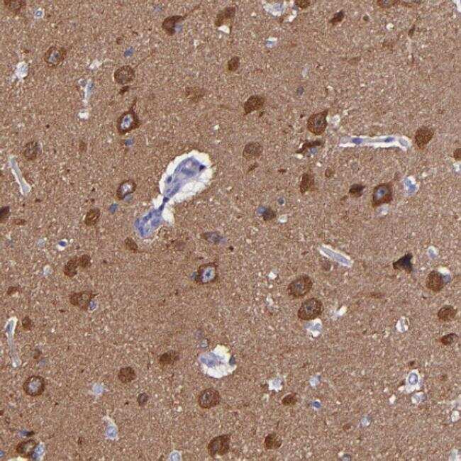
- Immunohistochemical staining of RAP1GAP in human hippocampus shows cytoplasmic positivity in neuronal cells. Samples were probed using a RAP1GAP Polyclonal Antibody (Product # PA5-51662).
- Submitted by
- Invitrogen Antibodies (provider)
- Main image

- Experimental details
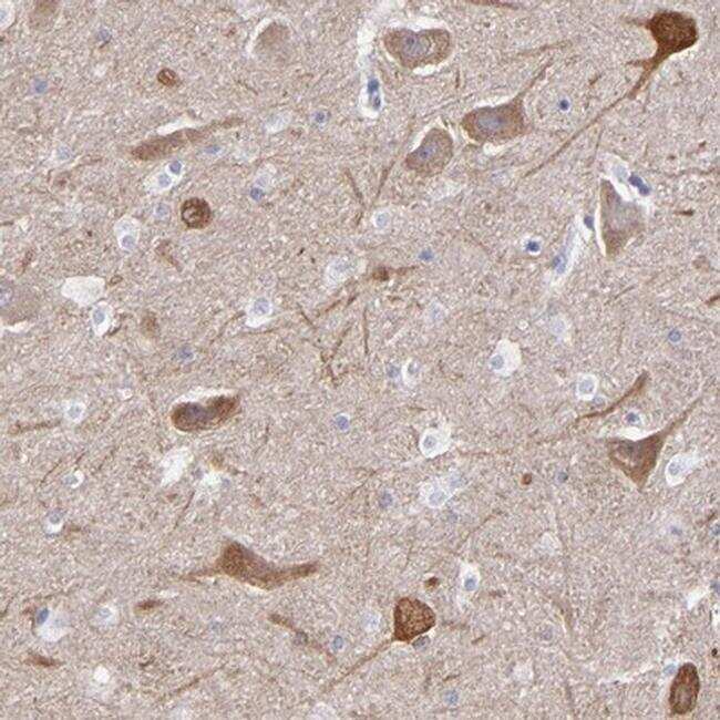
- Immunohistochemical staining of RAP1GAP in human cerebral cortex tissue shows cytoplasmic immunoreactivity in neuronal cell bodies. Samples were probed using a RAP1GAP Polyclonal Antibody (Product # PA5-51662).
- Submitted by
- Invitrogen Antibodies (provider)
- Main image

- Experimental details
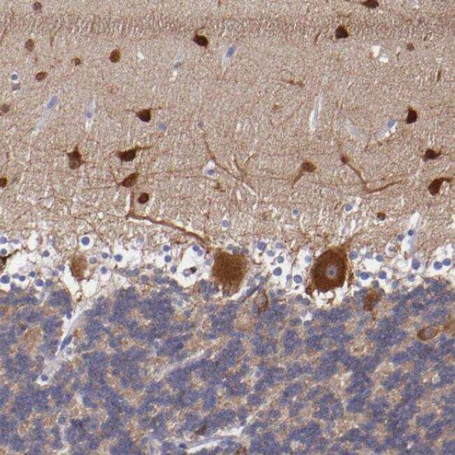
- Immunohistochemical staining of RAP1GAP in human cerebellum tissue shows strong cytoplasmic immunoreactivity in Purkinje cells and neurons in the molecular layer. Samples were probed using a RAP1GAP Polyclonal Antibody (Product # PA5-51662).
- Submitted by
- Invitrogen Antibodies (provider)
- Main image
- Experimental details
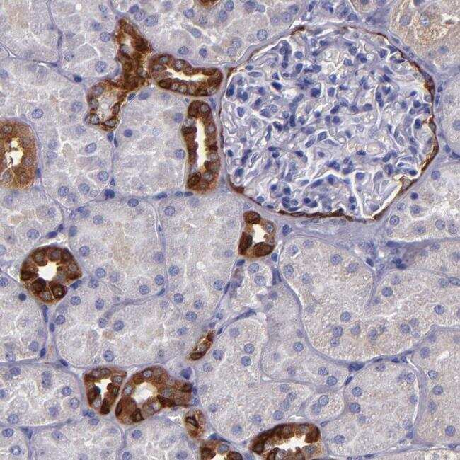
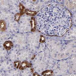
- Immunohistochemical staining of RAP1GAP in human kidney tissue shows strong positivity in distal tubules and Bowman's capsule. Samples were probed using a RAP1GAP Polyclonal Antibody (Product # PA5-51662).