 Explore
 Validate
 Learn
Learn Immunocytochemistry
Antibody data
- Antibody Data
- Antigen structure
- References [6]
- Comments [0]
- Validations
- Immunocytochemistry [1]
- Immunohistochemistry [1]
Submit
Validation data
Reference
Comment
Report error
- Product number
- HPA001923 - Provider product page

- Provider
- Atlas Antibodies
- Proper citation
- Atlas Antibodies Cat#HPA001923, RRID:AB_1080065
- Product name
- Anti-SOX6
- Antibody type
- Polyclonal
- Description
- Polyclonal Antibody against Human SOX6, Gene description: SRY (sex determining region Y)-box 6, Validated applications: ICC, IHC, Uniprot ID: P35712, Storage: Store at +4°C for short term storage. Long time storage is recommended at -20°C.
- Reactivity
- Human
- Host
- Rabbit
- Conjugate
- Unconjugated
- Isotype
- IgG
- Vial size
- 100 µl
- Concentration
- 0.1 mg/ml
- Storage
- Store at +4°C for short term storage. Long time storage is recommended at -20°C.
- Handling
- The antibody solution should be gently mixed before use.
Submitted references Sox6 expression distinguishes dorsally and ventrally biased dopamine neurons in the substantia nigra with distinctive properties and embryonic origins
Disruption of Interneuron Neurogenesis in Premature Newborns and Reversal with Estrogen Treatment
Estrogen Treatment Reverses Prematurity-Induced Disruption in Cortical Interneuron Population
Similar Properties of Chondrocytes from Osteoarthritis Joints and Mesenchymal Stem Cells from Healthy Donors for Tissue Engineering of Articular Cartilage
Immunofluorescence and fluorescent-protein tagging show high correlation for protein localization in mammalian cells
Chondrogenic Differentiation of Human Bone Marrow-Derived Mesenchymal Stem Cells in Self-Gelling Alginate Discs Reveals Novel Chondrogenic Signature Gene Clusters
Pereira Luppi M, Azcorra M, Caronia-Brown G, Poulin J, Gaertner Z, Gatica S, Moreno-Ramos O, Nouri N, Dubois M, Ma Y, Ramakrishnan C, Fenno L, Kim Y, Deisseroth K, Cicchetti F, Dombeck D, Awatramani R
Cell Reports 2021;37(6):109975
Cell Reports 2021;37(6):109975
Disruption of Interneuron Neurogenesis in Premature Newborns and Reversal with Estrogen Treatment
Tibrewal M, Cheng B, Dohare P, Hu F, Mehdizadeh R, Wang P, Zheng D, Ungvari Z, Ballabh P
The Journal of Neuroscience 2018;38(5):1100-1113
The Journal of Neuroscience 2018;38(5):1100-1113
Estrogen Treatment Reverses Prematurity-Induced Disruption in Cortical Interneuron Population
Panda S, Dohare P, Jain S, Parikh N, Singla P, Mehdizadeh R, Klebe D, Kleinman G, Cheng B, Ballabh P
The Journal of Neuroscience 2018;38(34):7378-7391
The Journal of Neuroscience 2018;38(34):7378-7391
Similar Properties of Chondrocytes from Osteoarthritis Joints and Mesenchymal Stem Cells from Healthy Donors for Tissue Engineering of Articular Cartilage
Burns J, Fernandes A, Herlofsen S, Karlsen T, Küchler A, Fløisand Y, Brinchmann J
PLoS ONE 2013;8(5):e62994
PLoS ONE 2013;8(5):e62994
Immunofluorescence and fluorescent-protein tagging show high correlation for protein localization in mammalian cells
Stadler C, Rexhepaj E, Singan V, Murphy R, Pepperkok R, Uhlén M, Simpson J, Lundberg E
Nature Methods 2013;10(4):315-323
Nature Methods 2013;10(4):315-323
Chondrogenic Differentiation of Human Bone Marrow-Derived Mesenchymal Stem Cells in Self-Gelling Alginate Discs Reveals Novel Chondrogenic Signature Gene Clusters
Herlofsen S, Küchler A, Melvik J, Brinchmann J
Tissue Engineering Part A 2011;17(7-8):1003-1013
Tissue Engineering Part A 2011;17(7-8):1003-1013
No comments: Submit comment
Supportive validation
- Submitted by
- Atlas Antibodies (provider)
- Main image

- Experimental details
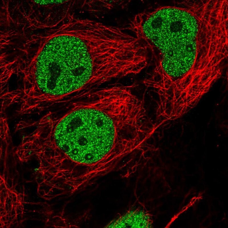
- Immunofluorescent staining of human cell line CACO-2 shows localization to nucleoplasm.
- Sample type
- Human
Supportive validation
- Submitted by
- Atlas Antibodies (provider)
- Enhanced method
- Orthogonal validation
- Main image
- Experimental details
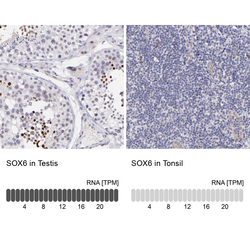
- Immunohistochemistry analysis in human testis and tonsil tissues using HPA001923 antibody. Corresponding SOX6 RNA-seq data are presented for the same tissues.
- Sample type
- Human
- Protocol
- Protocol
