Explore
 Validate
 Learn
Learn Western blot
 Immunocytochemistry
Antibody data
- Antibody Data
- Antigen structure
- References [3]
- Comments [0]
- Validations
- Immunocytochemistry [1]
Submit
Validation data
Reference
Comment
Report error
- Product number
- HPA001925 - Provider product page

- Provider
- Atlas Antibodies
- Proper citation
- Atlas Antibodies Cat#HPA001925, RRID:AB_1857756
- Product name
- Anti-TAGLN2
- Antibody type
- Polyclonal
- Description
- Polyclonal Antibody against Human TAGLN2, Gene description: transgelin 2, Alternative Gene Names: HA1756, KIAA0120, Validated applications: WB, IHC, ICC, Uniprot ID: P37802, Storage: Store at +4°C for short term storage. Long time storage is recommended at -20°C.
- Reactivity
- Human, Mouse, Rat
- Host
- Rabbit
- Conjugate
- Unconjugated
- Isotype
- IgG
- Vial size
- 100 µl
- Concentration
- 0.1 mg/ml
- Storage
- Store at +4°C for short term storage. Long time storage is recommended at -20°C.
- Handling
- The antibody solution should be gently mixed before use.
Submitted references Systematic validation of antibody binding and protein subcellular localization using siRNA and confocal microscopy
miR-1 as a tumor suppressive microRNA targeting TAGLN2 in head and neck squamous cell carcinoma
The tumour-suppressive function of miR-1 and miR-133a targeting TAGLN2 in bladder cancer
Stadler C, Hjelmare M, Neumann B, Jonasson K, Pepperkok R, Uhlén M, Lundberg E
Journal of Proteomics 2012;75(7):2236-2251
Journal of Proteomics 2012;75(7):2236-2251
miR-1 as a tumor suppressive microRNA targeting TAGLN2 in head and neck squamous cell carcinoma
Nohata N, Sone Y, Hanazawa T, Fuse M, Kikkawa N, Yoshino H, Chiyomaru T, Kawakami K, Enokida H, Nakagawa M, Shozu M, Okamoto Y, Seki N
Oncotarget 2011;2(1-2):29-42
Oncotarget 2011;2(1-2):29-42
The tumour-suppressive function of miR-1 and miR-133a targeting TAGLN2 in bladder cancer
Yoshino H, Chiyomaru T, Enokida H, Kawakami K, Tatarano S, Nishiyama K, Nohata N, Seki N, Nakagawa M
British Journal of Cancer 2011;104(5):808-818
British Journal of Cancer 2011;104(5):808-818
No comments: Submit comment
Supportive validation
- Submitted by
- Atlas Antibodies (provider)
- Main image
- Experimental details
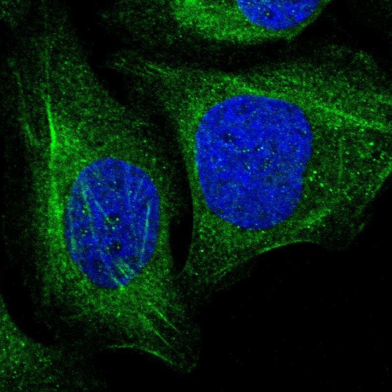
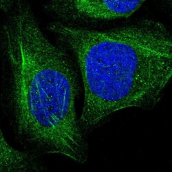
- Immunofluorescent staining of human cell line U-2 OS shows localization to cytosol & actin filaments.
- Sample type
- Human