 Explore
 Validate
 Learn
Learn Western blot
 Immunocytochemistry
Antibody data
- Antibody Data
- Antigen structure
- References [13]
- Comments [0]
- Validations
- Immunocytochemistry [1]
Submit
Validation data
Reference
Comment
Report error
- Product number
- HPA001927 - Provider product page

- Provider
- Atlas Antibodies
- Proper citation
- Atlas Antibodies Cat#HPA001927, RRID:AB_1079674
- Product name
- Anti-PREX1
- Antibody type
- Polyclonal
- Description
- Polyclonal Antibody against Human PREX1, Gene description: phosphatidylinositol-3,4,5-trisphosphate-dependent Rac exchange factor 1, Alternative Gene Names: KIAA1415, P-REX1, Validated applications: ICC, IHC, WB, Uniprot ID: Q8TCU6, Storage: Store at +4°C for short term storage. Long time storage is recommended at -20°C.
- Reactivity
- Human
- Host
- Rabbit
- Conjugate
- Unconjugated
- Isotype
- IgG
- Vial size
- 100 µl
- Concentration
- 0.2 mg/ml
- Storage
- Store at +4°C for short term storage. Long time storage is recommended at -20°C.
- Handling
- The antibody solution should be gently mixed before use.
Submitted references Migration, invasion, and metastasis are mediated by P-Rex1 in neuroblastoma
A cross-species proteomic map reveals neoteny of human synapse development
The pseudokinase NRBP1 activates Rac1/Cdc42 via P-Rex1 to drive oncogenic signalling in triple-negative breast cancer
cAMP-dependent activation of the Rac guanine exchange factor P-REX1 by type I protein kinase A (PKA) regulatory subunits
Gβγ signaling to the chemotactic effector P-REX1 and mammalian cell migration is directly regulated by Gαq and Gα13 proteins
Protumoral bone marrow-derived cells migrate via Gβγ-dependent signaling pathways and exhibit a complex repertoire of RhoGEFs
Zeb1 potentiates genome‐wide gene transcription with Lef1 to promote glioblastoma cell invasion
P-Rex1 Expression in Invasive Breast Cancer in relation to Receptor Status and Distant Metastatic Site
Differential Rac1 signalling by guanine nucleotide exchange factors implicates FLII in regulating Rac1-driven cell migration
Protein Kinase A (PKA) Type I Interacts with P-Rex1, a Rac Guanine Nucleotide Exchange Factor
P-Rex1 Cooperates with PDGFRβ to Drive Cellular Migration in 3D Microenvironments
Immunofluorescence and fluorescent-protein tagging show high correlation for protein localization in mammalian cells
P-Rex1 is required for efficient melanoblast migration and melanoma metastasis
Jacobson J, Qiao J, Cochran E, McCreery S, Chung D
Frontiers in Oncology 2024;14
Frontiers in Oncology 2024;14
A cross-species proteomic map reveals neoteny of human synapse development
Wang L, Pang K, Zhou L, Cebrián-Silla A, González-Granero S, Wang S, Bi Q, White M, Ho B, Li J, Li T, Perez Y, Huang E, Winkler E, Paredes M, Kovner R, Sestan N, Pollen A, Liu P, Li J, Piao X, García-Verdugo J, Alvarez-Buylla A, Liu Z, Kriegstein A
Nature 2023;622(7981):112-119
Nature 2023;622(7981):112-119
The pseudokinase NRBP1 activates Rac1/Cdc42 via P-Rex1 to drive oncogenic signalling in triple-negative breast cancer
Yang X, Cruz M, Nguyen E, Huang C, Schittenhelm R, Luu J, Cowley K, Shin S, Nguyen L, Lim Kam Sian T, Clark K, Simpson K, Ma X, Daly R
Oncogene 2023;42(11):833-847
Oncogene 2023;42(11):833-847
cAMP-dependent activation of the Rac guanine exchange factor P-REX1 by type I protein kinase A (PKA) regulatory subunits
Adame-García S, Cervantes-Villagrana R, Orduña-Castillo L, del Rio J, Gutkind J, Reyes-Cruz G, Taylor S, Vázquez-Prado J
Journal of Biological Chemistry 2019;294(7):2232-2246
Journal of Biological Chemistry 2019;294(7):2232-2246
Gβγ signaling to the chemotactic effector P-REX1 and mammalian cell migration is directly regulated by Gαq and Gα13 proteins
Cervantes-Villagrana R, Adame-García S, García-Jiménez I, Color-Aparicio V, Beltrán-Navarro Y, König G, Kostenis E, Reyes-Cruz G, Gutkind J, Vázquez-Prado J
Journal of Biological Chemistry 2019;294(2):531-546
Journal of Biological Chemistry 2019;294(2):531-546
Protumoral bone marrow-derived cells migrate via Gβγ-dependent signaling pathways and exhibit a complex repertoire of RhoGEFs
Cervantes-Villagrana R, Color-Aparicio V, Reyes-Cruz G, Vázquez-Prado J
Journal of Cell Communication and Signaling 2019;13(2):179-191
Journal of Cell Communication and Signaling 2019;13(2):179-191
Zeb1 potentiates genome‐wide gene transcription with Lef1 to promote glioblastoma cell invasion
Rosmaninho P, Mükusch S, Piscopo V, Teixeira V, Raposo A, Warta R, Bennewitz R, Tang Y, Herold‐Mende C, Stifani S, Momma S, Castro D
The EMBO Journal 2018;37(15)
The EMBO Journal 2018;37(15)
P-Rex1 Expression in Invasive Breast Cancer in relation to Receptor Status and Distant Metastatic Site
Marotti J, Muller K, Tafe L, Demidenko E, Miller T
International Journal of Breast Cancer 2017;2017
International Journal of Breast Cancer 2017;2017
Differential Rac1 signalling by guanine nucleotide exchange factors implicates FLII in regulating Rac1-driven cell migration
Marei H, Carpy A, Woroniuk A, Vennin C, White G, Timpson P, Macek B, Malliri A
Nature Communications 2016;7(1)
Nature Communications 2016;7(1)
Protein Kinase A (PKA) Type I Interacts with P-Rex1, a Rac Guanine Nucleotide Exchange Factor
Chávez-Vargas L, Adame-García S, Cervantes-Villagrana R, Castillo-Kauil A, Bruystens J, Fukuhara S, Taylor S, Mochizuki N, Reyes-Cruz G, Vázquez-Prado J
Journal of Biological Chemistry 2016;291(12):6182-6199
Journal of Biological Chemistry 2016;291(12):6182-6199
P-Rex1 Cooperates with PDGFRβ to Drive Cellular Migration in 3D Microenvironments
Facchiano A, Campbell A, Lawn S, McGarry L, Welch H, Ozanne B, Norman J
PLoS ONE 2013;8(1):e53982
PLoS ONE 2013;8(1):e53982
Immunofluorescence and fluorescent-protein tagging show high correlation for protein localization in mammalian cells
Stadler C, Rexhepaj E, Singan V, Murphy R, Pepperkok R, Uhlén M, Simpson J, Lundberg E
Nature Methods 2013;10(4):315-323
Nature Methods 2013;10(4):315-323
P-Rex1 is required for efficient melanoblast migration and melanoma metastasis
Lindsay C, Lawn S, Campbell A, Faller W, Rambow F, Mort R, Timpson P, Li A, Cammareri P, Ridgway R, Morton J, Doyle B, Hegarty S, Rafferty M, Murphy I, McDermott E, Sheahan K, Pedone K, Finn A, Groben P, Thomas N, Hao H, Carson C, Norman J, Machesky L, Gallagher W, Jackson I, Van Kempen L, Beermann F, Der C, Larue L, Welch H, Ozanne B, Sansom O
Nature Communications 2011;2(1)
Nature Communications 2011;2(1)
No comments: Submit comment
Supportive validation
- Submitted by
- Atlas Antibodies (provider)
- Main image
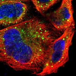
- Experimental details
- Immunofluorescent staining of human cell line A-431 shows localization to cytosol & vesicles.
- Sample type
- Human
