Explore
 Validate
 Learn
LearnAP7636a
antibody from Abcepta
Targeting: FGFR1
BFGFR, CD331, CEK, FLG, FLT2, H2, H3, H4, H5, KAL2, N-SAM
 Western blot
Antibody data
- Antibody Data
- Antigen structure
- References [4]
- Comments [0]
- Validations
- Western blot [1]
- Immunocytochemistry [1]
- Immunohistochemistry [1]
- Flow cytometry [1]
Submit
Validation data
Reference
Comment
Report error
- Product number
- AP7636a - Provider product page

- Provider
- Abcepta
- Proper citation
- Abgent Cat#AP7636A, RRID:AB_2293928
- Product name
- FGFR1 Antibody (N-term)
- Antibody type
- Polyclonal
- Antigen
- Synthetic peptide
- Description
- Purified Rabbit Polyclonal Antibody (Pab)
- Reactivity
- Human, Mouse
- Host
- Rabbit
- Isotype
- IgG
- Vial size
- 400 µl
- Concentration
- 2 mg/ml
- Storage
- Maintain refrigerated at 2-8°C for up to 6 months. For long term storage store at -20°C in small aliquots to prevent freeze-thaw cycles.
Submitted references L1CAM stimulates glioma cell motility and proliferation through the fibroblast growth factor receptor.
Induction of stem cell gene expression in adult human fibroblasts without transgenes.
Stimulation of endothelial cell proliferation by FGF-2 in the presence of fibrinogen requires alphavbeta3.
[Expeditions to high altitudes--what can we learn from them?].
Mohanan V, Temburni MK, Kappes JC, Galileo DS
Clinical & experimental metastasis 2013 Apr;30(4):507-20
Clinical & experimental metastasis 2013 Apr;30(4):507-20
Induction of stem cell gene expression in adult human fibroblasts without transgenes.
Page RL, Ambady S, Holmes WF, Vilner L, Kole D, Kashpur O, Huntress V, Vojtic I, Whitton H, Dominko T
Cloning and stem cells 2009 Sep;11(3):417-26
Cloning and stem cells 2009 Sep;11(3):417-26
Stimulation of endothelial cell proliferation by FGF-2 in the presence of fibrinogen requires alphavbeta3.
Sahni A, Francis CW
Blood 2004 Dec 1;104(12):3635-41
Blood 2004 Dec 1;104(12):3635-41
[Expeditions to high altitudes--what can we learn from them?].
Hauge A
Tidsskrift for den Norske laegeforening : tidsskrift for praktisk medicin, ny raekke 1991 Mar 20;111(8):926-8
Tidsskrift for den Norske laegeforening : tidsskrift for praktisk medicin, ny raekke 1991 Mar 20;111(8):926-8
No comments: Submit comment
Supportive validation
- Submitted by
- Abcepta (provider)
- Main image
- Experimental details
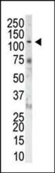
- The anti-FGFR1 Pab (Cat. #AP7636a) is used in Western blot to detect FGFR1 in NIH-3T3 cell lysate.
- Primary Ab dilution
- 1:1000
Supportive validation
- Submitted by
- Abcepta (provider)
- Main image
- Experimental details
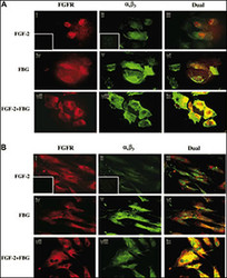
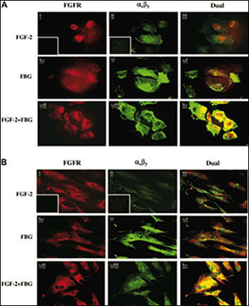
- "Colocalization of A1B3 and FGFR1 using IF. Confluent ECs (A) or HFFs (B) were treated with or without 100 ng/mL FGF-2 in the presence or absence of 10/mL fibrinogen. After 1 hour, cells were washed and fixed with 3.7% formaldehyde and stained using 10/mL FGFR1 and 7E3 antibody. FGFR is visualized as red fluorescence (i,iv,vii), A1B3 is visualized as green fluorescence (ii,v,viii), and colocalization of FGF-2 and fibrinogen receptors is shown as yellow fluorescence (iii,vi,ix). Insets represent the background staining for red (i) and green (ii) fluorescence. Bars represent 25 ."
- Primary Ab dilution
- 1:50~100
Supportive validation
- Submitted by
- Abcepta (provider)
- Main image
- Experimental details
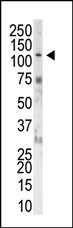
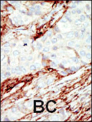
- "Formalin-fixed and paraffin-embedded human cancer tissue reacted with the primary antibody, which was peroxidase-conjugated to the secondary antibody, followed by DAB staining. This data demonstrates the use of this antibody for immunohistochemistry; clinical relevance has not been evaluated. BC = breast carcinoma; HC = hepatocarcinoma."
- Primary Ab dilution
- 1:50~100

Supportive validation
- Submitted by
- Abcepta (provider)
- Main image
- Experimental details
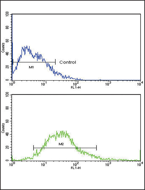
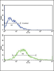
- Flow cytometric analysis of MCF-7 cells using FGFR1 Antibody (N-term) (bottom histogram) compared to a negative control cell (top histogram). FITC-conjugated goat-anti-rabbit secondary antibodies were used for the analysis.
- Primary Ab dilution
- 1:10~50