 Explore
 Validate
 Learn
Learn13-3100
antibody from Invitrogen Antibodies
Targeting: FGFR1
BFGFR, CD331, CEK, FLG, FLT2, H2, H3, H4, H5, KAL2, N-SAM
 Western blot
 Immunohistochemistry
Antibody data
- Antibody Data
- Antigen structure
- References [5]
- Comments [0]
- Validations
- Immunohistochemistry [1]
- Flow cytometry [1]
- Other assay [1]
Submit
Validation data
Reference
Comment
Report error
- Product number
- 13-3100 - Provider product page

- Provider
- Invitrogen Antibodies
- Product name
- FGFR1 Monoclonal Antibody (VBS-7)
- Antibody type
- Monoclonal
- Antigen
- Purifed from natural sources
- Reactivity
- Human, Mouse, Rat, Bovine, Chicken/Avian, Guinea Pig
- Host
- Mouse
- Isotype
- IgG
- Antibody clone number
- VBS-7
- Vial size
- 100 μg
- Concentration
- 0.5 mg/mL
- Storage
- -20°C
Submitted references Acidic fibroblast growth factor underlies microenvironmental regulation of MYC in pancreatic cancer.
A quantitative proteomic analysis uncovers the relevance of CUL3 in bladder cancer aggressiveness.
L1CAM stimulates glioma cell motility and proliferation through the fibroblast growth factor receptor.
Fragile histidine triad expression delays tumor development and induces apoptosis in human pancreatic cancer.
Fibroblast growth factor and epidermal growth factor receptors in ligament healing.
Bhattacharyya S, Oon C, Kothari A, Horton W, Link J, Sears RC, Sherman MH
The Journal of experimental medicine 2020 Aug 3;217(8)
The Journal of experimental medicine 2020 Aug 3;217(8)
A quantitative proteomic analysis uncovers the relevance of CUL3 in bladder cancer aggressiveness.
Grau L, Luque-Garcia JL, González-Peramato P, Theodorescu D, Palou J, Fernandez-Gomez JM, Sánchez-Carbayo M
PloS one 2013;8(1):e53328
PloS one 2013;8(1):e53328
L1CAM stimulates glioma cell motility and proliferation through the fibroblast growth factor receptor.
Mohanan V, Temburni MK, Kappes JC, Galileo DS
Clinical & experimental metastasis 2013 Apr;30(4):507-20
Clinical & experimental metastasis 2013 Apr;30(4):507-20
Fragile histidine triad expression delays tumor development and induces apoptosis in human pancreatic cancer.
Dumon KR, Ishii H, Vecchione A, Trapasso F, Baldassarre G, Chakrani F, Druck T, Rosato EF, Williams NN, Baffa R, During MJ, Huebner K, Croce CM
Cancer research 2001 Jun 15;61(12):4827-36
Cancer research 2001 Jun 15;61(12):4827-36
Fibroblast growth factor and epidermal growth factor receptors in ligament healing.
Panossian V, Liu SH, Lane JM, Finerman GA
Clinical orthopaedics and related research 1997 Sep;(342):173-80
Clinical orthopaedics and related research 1997 Sep;(342):173-80
No comments: Submit comment
Supportive validation
- Submitted by
- Invitrogen Antibodies (provider)
- Main image
- Experimental details
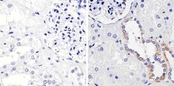
- Immunohistochemistry analysis of FGFR1/CD331 showing staining in the cytoplasm and membrane of paraffin-embedded human kidney tissue (right) compared to a negative control without primary antibody (left). To expose target proteins, antigen retrieval was performed using 10mM sodium citrate (pH 6.0), microwaved for 8-15 min. Following antigen retrieval, tissues were blocked in 3% H2O2-methanol for 15 min at room temperature, washed with ddH2O and PBS, and then probed with a FGFR1/CD331 monoclonal antibody (Product # 13-3100) diluted in 3% BSA-PBS at a dilution of 1:100 overnight at 4ºC in a humidified chamber. Tissues were washed extensively in PBST and detection was performed using an HRP-conjugated secondary antibody followed by colorimetric detection using a DAB kit. Tissues were counterstained with hematoxylin and dehydrated with ethanol and xylene to prep for mounting.

Supportive validation
- Submitted by
- Invitrogen Antibodies (provider)
- Main image

- Experimental details
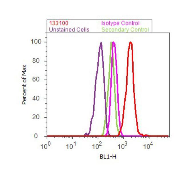
- Flow cytometry analysis of FGFR1 was done on SH-SY5Y cells treated with FGF (100ng/mL, 20 minutes). Cells were fixed with 70% ethanol for 10 minutes, permeabilized with 0.25% Triton™ X-100 for 20 minutes, and blocked with 5% BSA for 30 minutes at room temperature. Cells were labeled with FGFR1 Mouse Monoclonal Antibody (133100, red histogram) or with mouse isotype control (pink histogram) at 3-5 ug/million cells in 2.5% BSA. After incubation at room temperature for 2 hours, the cells were labeled with Alexa Fluor® 488 Rabbit Anti-Mouse Secondary Antibody (A11059) at a dilution of 1:400 for 30 minutes at room temperature. The representative 10,000 cells were acquired and analyzed for each sample using an Attune® Acoustic Focusing Cytometer. The purple histogram represents unstained control cells and the green histogram represents no-primary-antibody control.
Supportive validation
- Submitted by
- Invitrogen Antibodies (provider)
- Main image
- Experimental details
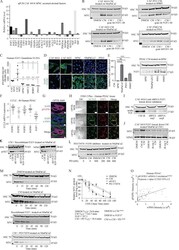
- Figure 2. Stroma-derived FGF1 is necessary and sufficient for paracrine regulation of MYC. (A) qRT-PCR for the indicated secreted factors in CAF 4414 and hPSC ( n = 3 independent experiments). Data are plotted as fold change in CAF 4414 compared with hPSC (set to 1); all values were normalized to 36B4 as a housekeeping gene. ( B) CAF CM was incubated alone or with FGF1 neutralizing antibody or goat IgG control antibody for 1 h at room temperature, then added to PDAC cells for 3 h. MYC levels in nuclear extracts were analyzed by Western blot (representative of three independent experiments). ( C) ELISA for FGF1 in CM from the indicated cell lines. Data are presented as mean +- SEM; *, P < 0.05; **, P < 0.0025; ***, P < 0.001; ****, P < 0.0001 by Student's t test ( n = 3 independent experiments) compared with CAF 4422 CM (upper line and asterisks) or CAF 4414 CM (lower line and asterisks). (D) IF staining for FGF1 or FGFR1 in the indicated cell lines. Scale bar, 10 um. (E) hPSCs were treated with CM from the indicated PDAC cell lines for 24 h, and FGF1 mRNA was measured by qPCR (left; data were normalized to 36B4 and are presented as mean +- SEM; ***, P < 0.001; ****, P < 0.0001 by Student's t test) and protein measured by Western blot (right). Results represent three independent experiments. (F) mRNA expression levels of the four FGFRs and FGF1 in 40 human PDAC cells from the Cancer Cell Line Encyclopedia database. (G) Fluorescent immunostaining for pFGFR1 in human PDAC sample
