Explore
 Validate
 Learn
LearnAF3285
antibody from R&D Systems
Targeting: FGFR1
BFGFR, CD331, CEK, FLG, FLT2, H2, H3, H4, H5, KAL2, N-SAM
 Western blot
Antibody data
- Antibody Data
- Antigen structure
- References [4]
- Comments [0]
- Validations
- Western blot [1]
- Immunocytochemistry [1]
Submit
Validation data
Reference
Comment
Report error
- Product number
- AF3285 - Provider product page

- Provider
- R&D Systems
- Product name
- Human Phospho-FGFR1-4 (Y653/Y654) Antibody
- Antibody type
- Polyclonal
- Description
- Antigen Affinity-purified. Detects human FGF R when dually phosphorylated at Y653/Y654 in Western blots.
- Reactivity
- Human
- Host
- Rabbit
- Conjugate
- Unconjugated
- Isotype
- IgG
- Vial size
- 50 ug
- Concentration
- LYOPH
- Storage
- Use a manual defrost freezer and avoid repeated freeze-thaw cycles. 12 months from date of receipt, -20 to -70 °C as supplied. 1 month, 2 to 8 °C under sterile conditions after reconstitution. 6 months, -20 to -70 °C under sterile conditions after reconstitution.
Submitted references FGFR2 amplification is predictive of sensitivity to regorafenib in gastric and colorectal cancers in vitro.
Gambogenic acid inhibits fibroblast growth factor receptor signaling pathway in erlotinib-resistant non-small-cell lung cancer and suppresses patient-derived xenograft growth.
Basic Fibroblast Growth Factor Inhibits Apoptosis and Promotes Proliferation of Adipose-Derived Mesenchymal Stromal Cells Isolated from Patients with Type 2 Diabetes by Reducing Cellular Oxidative Stress.
FGFR2-amplified gastric cancer cell lines require FGFR2 and Erbb3 signaling for growth and survival.
Cha Y, Kim HP, Lim Y, Han SW, Song SH, Kim TY
Molecular oncology 2018 Jun;12(7):993-1003
Molecular oncology 2018 Jun;12(7):993-1003
Gambogenic acid inhibits fibroblast growth factor receptor signaling pathway in erlotinib-resistant non-small-cell lung cancer and suppresses patient-derived xenograft growth.
Xu L, Meng X, Xu N, Fu W, Tan H, Zhang L, Zhou Q, Qian J, Tu S, Li X, Lao Y, Xu H
Cell death & disease 2018 Feb 15;9(3):262
Cell death & disease 2018 Feb 15;9(3):262
Basic Fibroblast Growth Factor Inhibits Apoptosis and Promotes Proliferation of Adipose-Derived Mesenchymal Stromal Cells Isolated from Patients with Type 2 Diabetes by Reducing Cellular Oxidative Stress.
Nawrocka D, Kornicka K, Szydlarska J, Marycz K
Oxidative medicine and cellular longevity 2017;2017:3027109
Oxidative medicine and cellular longevity 2017;2017:3027109
FGFR2-amplified gastric cancer cell lines require FGFR2 and Erbb3 signaling for growth and survival.
Kunii K, Davis L, Gorenstein J, Hatch H, Yashiro M, Di Bacco A, Elbi C, Lutterbach B
Cancer research 2008 Apr 1;68(7):2340-8
Cancer research 2008 Apr 1;68(7):2340-8
No comments: Submit comment
Supportive validation
- Submitted by
- R&D Systems (provider)
- Main image

- Experimental details
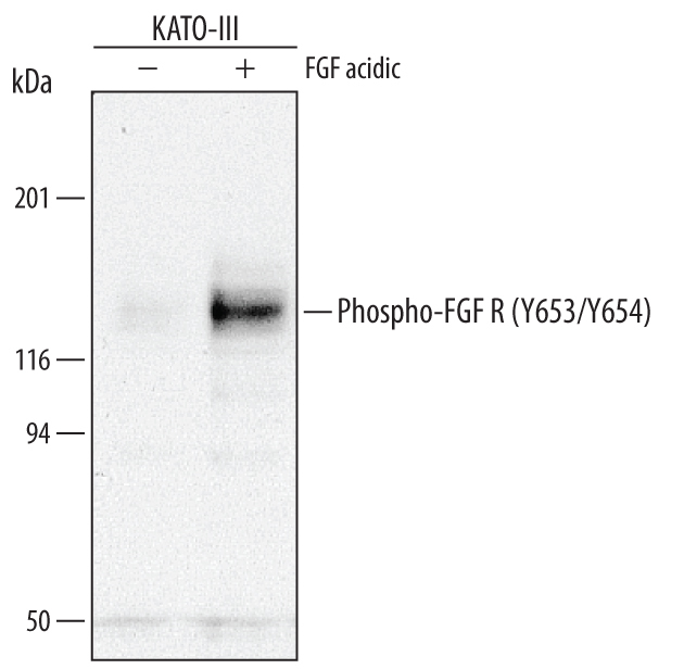
- Detection of Human Phospho- FGF R1-4 (Y653/Y654) by Western Blot. Western blot shows lysates of KATO-III human gastric carcinoma cell line untreated (-) or treated (+) with 100 ng/mL Recom-binant Human FGF acidic (Catalog # 232-FA) for 15 minutes. PVDF membrane was probed with 0.5 µg/mL of Rabbit Anti-Human Phospho-FGF R1-4 (Y653/Y654) Antigen Affinity-purified Polyclonal Antibody (Catalog # AF3285), followed by HRP-conjugated Anti-Rabbit IgG Secondary Antibody (Catalog # HAF008). A specific band was detected for Phospho-FGF R1-4 (Y653/Y654) at approximately 120 to 145 kDa (as indicated). This experiment was conducted under reducing conditions and using Immunoblot Buffer Group 1.
Supportive validation
- Submitted by
- R&D Systems (provider)
- Main image
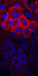
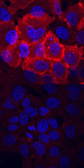
- Experimental details
- Phospho-FGF R1-4 (Y653/Y654) in A431 Human Cell Line. FGF R1-4 phosphorylated at Y653/Y654 was detected in immersion fixed A431 human epithelial carcinoma cell line untreated (lower panel) or treated (upper panel) with pervanadate using Rabbit Anti-Human Phospho-FGF R1-4 (Y653/Y654) Antigen Affinity-purified Polyclonal Antibody (Catalog # AF3285) at 10 µg/mL for 3 hours at room temperature. Cells were stained using the NorthernLights™ 557-conjugated Anti-Rabbit IgG Secondary Antibody (red; Catalog # NL004) and counterstained with DAPI (blue). View our protocol for Fluorescent ICC Staining of Cells on Coverslips.