Explore
 Validate
 Learn
LearnF50611
antibody from NSJ Bioreagents
Targeting: FGFR1
BFGFR, CD331, CEK, FLG, FLT2, H2, H3, H4, H5, KAL2, N-SAM
 Western blot
 ELISA
ELISAAntibody data
- Antibody Data
- Antigen structure
- References [0]
- Comments [0]
- Validations
- Western blot [1]
- Immunocytochemistry [1]
- Immunohistochemistry [1]
- Flow cytometry [1]
Submit
Validation data
Reference
Comment
Report error
- Product number
- F50611 - Provider product page

- Provider
- NSJ Bioreagents
- Product name
- FGFR1 Antibody
- Antibody type
- Polyclonal
- Description
- This highly specific FGFR1 antibody is suitable for use in Immunohistochemistry/Immunofluorescence/Flow cytometry/Western blot/ELISA applications with human and mouse samples.
- Reactivity
- Human, Mouse
- Host
- Rabbit
- Conjugate
- Unconjugated
- Vial size
- 0.08 ml, 0.4 ml
- Concentration
- In 1X PBS, pH 7.4, with 0.09% sodium azide
- Storage
- Aliquot the FGFR1 antibody and store frozen at -20oC or colder. Avoid repeated freeze-thaw cycles.
No comments: Submit comment
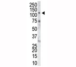
Supportive validation
- Submitted by
- NSJ Bioreagents (provider)
- Main image
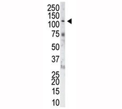
- Experimental details
- The FGFR1 antibody used in western blot to detect FGFR1 in NIH3T3 cell lysate. Predicted molecular weight: 75-160 kDa depending on glycosylation level.
Supportive validation
- Submitted by
- NSJ Bioreagents (provider)
- Main image
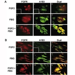
- Experimental details
- IF: Colocalization of A1B3 and FGFR1 using IF. Confluent ECs (A) or HFFs (B) were treated with or without 100 ng/mL FGF2 in the presence or absence of 10/mL fibrinogen. After 1 hour, cells were washed and fixed with 3.7% formaldehyde and stained using 7E3 and FGFR1 antibody. FGFR is visualized as red fluorescence (i,iv,vii), A1B3 is visualized as green fluorescence (ii,v,viii), and colocalization of FGF2 and fibrinogen receptors is shown as yellow fluorescence (iii,vi,ix). Insets represent the background fluorescence.

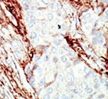
Supportive validation
- Submitted by
- NSJ Bioreagents (provider)
- Main image

- Experimental details
- IHC analysis of FFPE human breast carcinoma tissue stained with the FGFR1 antibody
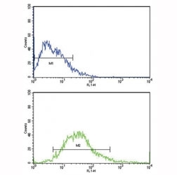
Supportive validation
- Submitted by
- NSJ Bioreagents (provider)
- Main image

- Experimental details
- Flow cytometric analysis of MCF-7 cells using FGFR1 antibody (bottom histogram) compared to a negative control (top histogram). FITC-conjugated goat-anti-rabbit secondary Ab was used for the analysis.