Explore
 Validate
 Learn
LearnNB100-2080
antibody from Novus Biologicals
Targeting: FGFR1
BFGFR, CD331, CEK, FLG, FLT2, H2, H3, H4, H5, KAL2, N-SAM
 Western blot
 Immunocytochemistry
Antibody data
- Antibody Data
- Antigen structure
- References [1]
- Comments [0]
- Validations
- Western blot [1]
- Immunohistochemistry [3]
- Flow cytometry [2]
Submit
Validation data
Reference
Comment
Report error
- Product number
- NB100-2080 - Provider product page

- Provider
- Novus Biologicals
- Proper citation
- Novus Cat#NB100-2080, RRID:AB_10002761
- Product name
- Mouse Monoclonal FGFR1 Antibody
- Antibody type
- Monoclonal
Submitted references Expression of a truncated FGF receptor results in defective lens development in transgenic mice.
Robinson ML, MacMillan-Crow LA, Thompson JA, Overbeek PA
Development (Cambridge, England) 1995 Dec;121(12):3959-67
Development (Cambridge, England) 1995 Dec;121(12):3959-67
No comments: Submit comment
Supportive validation
- Submitted by
- Novus Biologicals (provider)
- Main image
- Experimental details
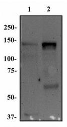
- Western Blot: FGF R1 Antibody (M17D10) [NB100-2080] - Total protein from MOLT-4 (lane 1) and Ntera-2 (lane 2) was separated on a 7.5% gel by SDS-PAGE. Protein was transferred to PVDF membrane, blocked and probed with 1 ug/mL of anti-FGF R1. FGF R1 protein was detected at ~150 kDa using an anti-mouse HRP secondary antibody.

Supportive validation
- Submitted by
- Novus Biologicals (provider)
- Main image
- Experimental details
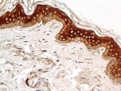
- Immunohistochemistry-Paraffin: FGF R1 Antibody (M17D10) [NB100-2080] - Analysis of FFPE tissue section of human normal skin using mouse monoclonal FGF R1 antibody (clone M17D10) at 5 ug/mL. Cells of the epidermal layer showed a very strong membrane-cytoplasmic staining with relatively weak nuclear immunopositivity for for this protein. The stratum basale / bottom layer of keratinocytes in the epidermis showed a membrane-cytoplasmic stainin for FGF R1.

- Submitted by
- Novus Biologicals (provider)
- Main image
- Experimental details
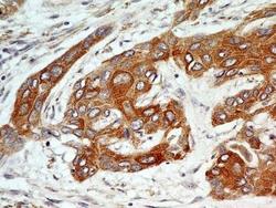
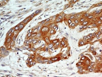
- Immunohistochemistry-Paraffin: FGF R1 Antibody (M17D10) [NB100-2080] - IHC analysis of formalin-fixed paraffin-embedded tissue section of human esophageal squamous cell carcinoma (SCC) using mouse monoclonal FGF R1 antibody (clone M17D10) at 5 ug/ml concentration. Specific membrane-cytoplasmic immunopositivity of FGF R1 protein was observed in SCC cells whereas the nuclei of SCC cells as well as the tumor stroma was largely negative for this protein.
- Submitted by
- Novus Biologicals (provider)
- Main image

- Experimental details
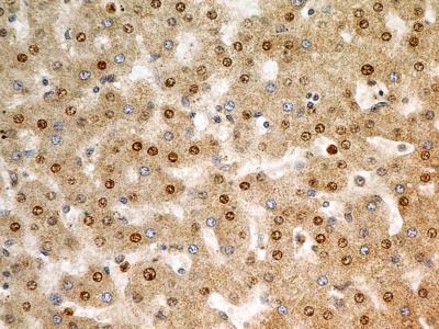
- Immunohistochemistry-Paraffin: FGF R1 Antibody (M17D10) [NB100-2080] - Analysis of FFPE tissue section of human liver using mouse monoclonal FGF R1 antibody (clone M17D10) at 5 ug/mL. The hepatocytes depicted an expected and specific membrane-cytoplasmic-nuclear immunostaining of FGF R1 protein.
Supportive validation
- Submitted by
- Novus Biologicals (provider)
- Main image
- Experimental details
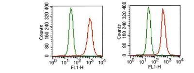
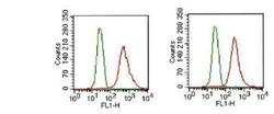
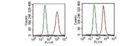
- Flow Cytometry: FGF R1 Antibody (M17D10) [NB100-2080] - Analysis of FGF R1 in MCF-7 cells (1x10^6 cells/mL) were stained with FGF R1 antibody (NB100-2080, red) at 1:1000. Detected with FITC conjugated goat anti-mouse IgG1 isotype control (green). Two distinct samples shown.
- Submitted by
- Novus Biologicals (provider)
- Main image
- Experimental details
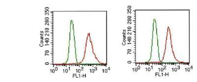
- Flow Cytometry: FGF R1 Antibody (M17D10) [NB100-2080] - Analysis of FGF R1 in HEK293 cells (2x10^6 cells/ml) were stained with FGF R1 antibody (NB100-2080, red) at 1:1000 dilution. Detected with FITC conjugated goat anti-mouse IgG1 isotype control (green). Two distinct samples shown.