Explore
 Validate
 Learn
LearnAMM85972
antibody from EnkiLife Biotech Co., Ltd.
Targeting: FGFR1
BFGFR, CD331, CEK, FLG, FLT2, H2, H3, H4, H5, KAL2, N-SAM
 Western blot
Antibody data
- Antibody Data
- Antigen structure
- References [0]
- Comments [0]
- Validations
- Western blot [1]
- Immunocytochemistry [1]
- Immunohistochemistry [1]
Submit
Validation data
Reference
Comment
Report error
- Product number
- AMM85972 - Provider product page

- Provider
- EnkiLife Biotech Co., Ltd.
- Product name
- FGFR1 Mouse Monoclonal Antibody
- Antibody type
- Monoclonal
- Description
- Affinity Purification
- Reactivity
- Human, Mouse
- Host
- Mouse
- Conjugate
- Unconjugated
- Antibody clone number
- Monoclonal
- Vial size
- 100 µl
- Concentration
- 1 mg/ml
- Storage
- Store at 4°C short term. Aliquot and store at -20°C long term. Avoid freeze/thaw cycles.
- Handling
- The antibody solution should be gently mixed before use.
No comments: Submit comment
Supportive validation
- Submitted by
- EnkiLife Biotech Co., Ltd. (provider)
- Main image
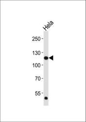
- Experimental details
- Western blot analysis of lysate from Hela cell line, using FGFR1 Antibody (C-term). FGFR1 Mouse Monoclonal Antibody was diluted at 1:2000. A goat anti-mouse IgG H&L(HRP) at 1:3000 dilution was used as the secondary antibody. Lysate at 20μg.

Supportive validation
- Submitted by
- EnkiLife Biotech Co., Ltd. (provider)
- Main image
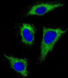
- Experimental details
- Immunofluorescent analysis of 4% paraformaldehyde-fixed, 0.1% Triton X-100 permeabilized HeLa (human cervical epithelial adenocarcinoma cell line) cells labeling FGFR1 with AMM85972 at 1/25 dilution, followed by Dylight® 488-conjugated goat anti-mouse IgG secondary antibody at 1/200 dilution (green). Immunofluorescence image showing cytoplasm staining on HeLa cell line. The nuclear counter stain is DAPI (blue).

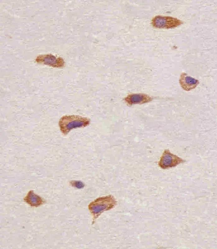
Supportive validation
- Submitted by
- EnkiLife Biotech Co., Ltd. (provider)
- Main image
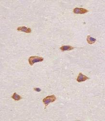
- Experimental details
- AMM85972 staining FGFR1 in human brain tissue sections by Immunohistochemistry (IHC-P - paraformaldehyde-fixed, paraffin-embedded sections). Tissue was fixed with formaldehyde and blocked with 3% BSA for 0. 5 hour at room temperature; antigen retrieval was by heat mediation with a citrate buffer (pH6). Samples were incubated with FGFR1 Mouse Monoclonal Antibody (1/25) for 1 hours at 37°C. A undiluted biotinylated goat polyvalent antibody was used as the secondary antibody.