Explore
 Validate
 Learn
Learn Western blot
 Immunohistochemistry
Antibody data
- Antibody Data
- Antigen structure
- References [6]
- Comments [0]
- Validations
- Immunohistochemistry [1]
Submit
Validation data
Reference
Comment
Report error
- Product number
- MAB669-100 - Provider product page

- Provider
- R&D Systems
- Product name
- Human Follistatin Antibody
- Antibody type
- Monoclonal
- Description
- Protein A or G purified from ascites. Detects human Follistatin in direct ELISAs and Western blots. It recognizes the 288 aa, 300 aa, and 315 aa isoforms of human Follistatin. In direct ELISAs and Western blots, this antibody shows approximately 50% cross-reactivity with recombinant mouse Follistatin.
- Reactivity
- Human
- Host
- Mouse
- Conjugate
- Unconjugated
- Antigen sequence
P19883- Isotype
- IgG
- Antibody clone number
- 85918
- Vial size
- 100 ug
- Storage
- Use a manual defrost freezer and avoid repeated freeze-thaw cycles. 12 months from date of receipt, -20 to -70 °C as supplied. 1 month, 2 to 8 °C under sterile conditions after reconstitution. 6 months, -20 to -70 °C under sterile conditions after reconstitution.
Submitted references Differentiation of nestin‑negative human hair follicle outer root sheath cells into neurons in vitro.
BMP-driven NRF2 activation in esophageal basal cell differentiation and eosinophilic esophagitis.
Nucleolar follistatin promotes cancer cell survival under glucose-deprived conditions through inhibiting cellular rRNA synthesis.
Canine follicle stem cell candidates reside in the bulge and share characteristic features with human bulge cells.
Thapsigargin resistance in human prostate cancer cells.
Differential gene expression and regulation of the bone morphogenetic protein antagonists follistatin and gremlin in normal and osteoarthritic human chondrocytes and synovial fibroblasts.
Wu W, Wu XL, Ji YQ, Gao Z
Molecular medicine reports 2017 Jul;16(1):95-100
Molecular medicine reports 2017 Jul;16(1):95-100
BMP-driven NRF2 activation in esophageal basal cell differentiation and eosinophilic esophagitis.
Jiang M, Ku WY, Zhou Z, Dellon ES, Falk GW, Nakagawa H, Wang ML, Liu K, Wang J, Katzka DA, Peters JH, Lan X, Que J
The Journal of clinical investigation 2015 Apr;125(4):1557-68
The Journal of clinical investigation 2015 Apr;125(4):1557-68
Nucleolar follistatin promotes cancer cell survival under glucose-deprived conditions through inhibiting cellular rRNA synthesis.
Gao X, Wei S, Lai K, Sheng J, Su J, Zhu J, Dong H, Hu H, Xu Z
The Journal of biological chemistry 2010 Nov 19;285(47):36857-64
The Journal of biological chemistry 2010 Nov 19;285(47):36857-64
Canine follicle stem cell candidates reside in the bulge and share characteristic features with human bulge cells.
Kobayashi T, Iwasaki T, Amagai M, Ohyama M
The Journal of investigative dermatology 2010 Aug;130(8):1988-95
The Journal of investigative dermatology 2010 Aug;130(8):1988-95
Thapsigargin resistance in human prostate cancer cells.
O'Neill JP, Velalar CN, Lee DI, Zhang B, Nakanishi T, Tang Y, Selaru F, Ross D, Meltzer SJ, Hussain A
Cancer 2006 Aug 1;107(3):649-59
Cancer 2006 Aug 1;107(3):649-59
Differential gene expression and regulation of the bone morphogenetic protein antagonists follistatin and gremlin in normal and osteoarthritic human chondrocytes and synovial fibroblasts.
Tardif G, Hum D, Pelletier JP, Boileau C, Ranger P, Martel-Pelletier J
Arthritis and rheumatism 2004 Aug;50(8):2521-30
Arthritis and rheumatism 2004 Aug;50(8):2521-30
No comments: Submit comment
Supportive validation
- Submitted by
- R&D Systems (provider)
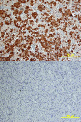
- Main image
- Experimental details
- Follistatin in Human Pituitary. Follistatin was detected in immersion fixed paraffin-embedded sections of human pituitary using Human Follistatin Monoclonal Antibody (Catalog # MAB669) at 25 µg/mL overnight at 4 °C. Tissue was stained using the Anti-Mouse HRP-DAB Cell & Tissue Staining Kit (brown; Catalog # CTS002) and counterstained with hematoxylin (blue). Lower panel shows a lack of labeling if primary antibodies are omitted and tissue is stained only with secondary antibody followed by incubation with detection reagents. View our protocol for Chromogenic IHC Staining of Paraffin-embedded Tissue Sections.