Explore
 Validate
 Learn
Learn Flow cytometry
Antibody data
- Antibody Data
- Antigen structure
- References [2]
- Comments [0]
- Validations
- Flow cytometry [1]
Submit
Validation data
Reference
Comment
Report error
- Product number
- 50-8897-42 - Provider product page

- Provider
- Invitrogen Antibodies
- Product name
- Granzyme K Monoclonal Antibody (G3H69), eFluor™ 660, eBioscience™
- Antibody type
- Monoclonal
- Antigen
- Other
- Description
- Description: This G3H69 monoclonal antibody reacts with human Granzyme K. Granzyme K is one of five granzyme serine proteases that have been indentified in humans. These proteins are expressed in the granules of NK cells and cytotoxic T cells, and are critical for the induction of target cell apoptosis through the cleavage of intracellular substrates. Granzyme A and Granzyme K are both tryptases and appear to show overlapping, though not identical, substrate specificity. This functional similarity is believed to account for the minimal decrease in cytotoxicity of Granzyme A-deficient CTLs. Applications Reported: This G3H69 antibody has been reported for use in intracellular staining followed by flow cytometric analysis. Applications Tested: This G3H69 antibody has been pre-titrated and tested by intracellular staining followed by flow cytometric analysis. This can be used at 5 µL (0.03 µg) per test. A test is defined as the amount (µg) of antibody that will stain a cell sample in a final volume of 100 µL. Cell number should be determined empirically but can range from 10^5 to 10^8 cells/test. eFluor® 660 is a replacement for Alexa Fluor® 647. eFluor® 660 emits at 659 nm and is excited with the red laser (633 nm). Please make sure that your instrument is capable of detecting this fluorochome. Excitation: 633-647 nm; Emission: 668 nm; Laser: Red Laser. Filtration: 0.2 µm post-manufacturing filtered.
- Reactivity
- Human
- Host
- Mouse
- Isotype
- IgG
- Antibody clone number
- G3H69
- Vial size
- 100 Tests
- Concentration
- 5 µL/Test
- Storage
- 4° C, store in dark, DO NOT FREEZE!
Submitted references Granzyme K displays highly restricted substrate specificity that only partially overlaps with granzyme A.
Residual cytotoxicity and granzyme K expression in granzyme A-deficient cytotoxic lymphocytes.
Bovenschen N, Quadir R, van den Berg AL, Brenkman AB, Vandenberghe I, Devreese B, Joore J, Kummer JA
The Journal of biological chemistry 2009 Feb 6;284(6):3504-12
The Journal of biological chemistry 2009 Feb 6;284(6):3504-12
Residual cytotoxicity and granzyme K expression in granzyme A-deficient cytotoxic lymphocytes.
Shresta S, Goda P, Wesselschmidt R, Ley TJ
The Journal of biological chemistry 1997 Aug 8;272(32):20236-44
The Journal of biological chemistry 1997 Aug 8;272(32):20236-44
No comments: Submit comment
Supportive validation
- Submitted by
- Invitrogen Antibodies (provider)
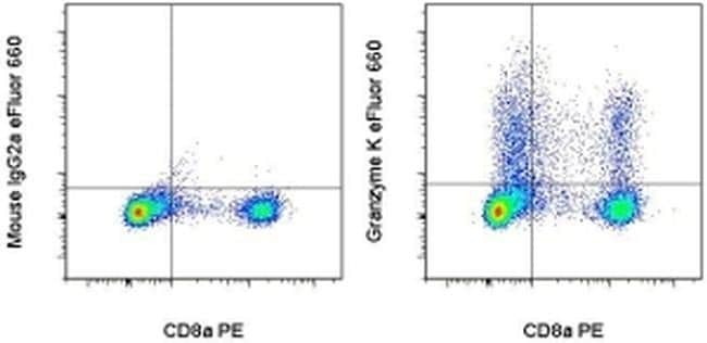
- Main image
- Experimental details
- Normal human peripheral blood monocytes were stimulated for 3 days with Human IL-2 Recombinant Protein (Product # 14-8029-81), then cultured with Protein Transport Inhibitor Cocktail (Product # 00-4980-03) for an additional 5 hours. The cells were stained with Anti-Human CD8a PE (Product # 12-0087-42) and Mouse IgG2a Isotype Control eFluor® 660 (Product # 50-4724-80) (left) or Anti-Human Granzyme K eFluor® 660 (right) using the Intracellular Fixation and Permeabilization Buffer Set (Product # 88-8824-00). Cells in the lymphocyte gate were used for analysis.