Explore
 Validate
 Learn
Learn Western blot
 ELISA
ELISAAntibody data
- Antibody Data
- Antigen structure
- References [0]
- Comments [0]
- Validations
- Western blot [2]
- Immunocytochemistry [1]
Submit
Validation data
Reference
Comment
Report error
- Product number
- 600-401-GU5 - Provider product page

- Provider
- Invitrogen Antibodies
- Product name
- PINK1 truncated Polyclonal Antibody
- Antibody type
- Polyclonal
- Antigen
- Synthetic peptide
- Reactivity
- Human, Mouse
- Host
- Rabbit
- Isotype
- IgG
- Vial size
- 100 µg
- Concentration
- 1 mg/mL
- Storage
- -20° C, Avoid Freeze/Thaw Cycles
No comments: Submit comment
Supportive validation
- Submitted by
- Invitrogen Antibodies (provider)
- Main image
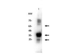
- Experimental details
- Western Blot of Rabbit anti-PINK1 truncated antibody. Lane 1: recombinant PINK1. Load: 1 µg per lane. Primary antibody: PINK1 truncated antibody at 1:1000 for overnight at 4°C. Secondary antibody: rabbit secondary HRP antibody p/n (611-103-122) at 1:70,000 for 45 min at RT. Block: Odyssey blocking buffer overnight at 4°C. Predicted/Observed size: ~62, 30/~30 kDa for PINK1. Other band(s): PINK1 phosphorylates and experiences rapid degradation causing various bands on WB.

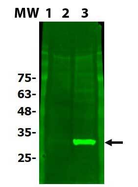
- Submitted by
- Invitrogen Antibodies (provider)
- Main image

- Experimental details
- Western Blot of Rabbit anti-PINK1 truncated antibody. Lane 1: MW ladder (opal pre-stained) p/n (MB-210-0500). Lane 2: HEK293T untransfected lysate (W09-001-GX5). Lane 3: PINK1-overexpressing 293T. Load: 10 µg per lane. Primary antibody: PINK1 truncated antibody at 1:1000 for overnight at 4°C. Secondary antibody: Anti-RABBIT IgG (H&L) (DONKEY) Antibody DyLight™ 488 Conjugated p/n (611-741-127) at 1:70,000 for 45 min at RT. Block: Odyssey blocking buffer overnight at 4°C. Predicted/Observed size: ~62, 30/~30 kDa for PINK1.
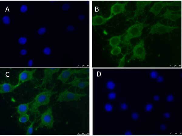
Supportive validation
- Submitted by
- Invitrogen Antibodies (provider)
- Main image
- Experimental details
- Immunofluorescence Microscopy of Rabbit anti-PINK1 truncated antibody. Cell line: NIH 3T3 (p/n W10-000-358). Fixation: 0.5% PFA. Antigen retrieval: not required. Primary antibody: PINK1 truncated antibody at 20 µg/mL for overnight at 4°C. Secondary antibody: Anti-RABBIT IgG DyLight™ 488 Conjugated Preadsorbed (p/n 611-741-127) at 5 µg/mL for 2 hrs at RT. Localization: PINK1 is mitochondrial and cytoplasmic. Staining: PINK1 as green fluorescent signal with DAPI (blue) nuclear counterstain.