Explore
 Validate
 Learn
Learn Flow cytometry
Antibody data
- Antibody Data
- Antigen structure
- References [3]
- Comments [0]
- Validations
- Flow cytometry [1]
- Other assay [1]
Submit
Validation data
Reference
Comment
Report error
- Product number
- 25-9177-41 - Provider product page

- Provider
- Invitrogen Antibodies
- Product name
- Granzyme A Monoclonal Antibody (CB9), PE-Cyanine7, eBioscience™
- Antibody type
- Monoclonal
- Antigen
- Other
- Description
- Description: This CB9 monoclonal antibody reacts with human Granzyme A. Granzyme A is the most abundantly expressed of the granzyme serine proteases, which are proteins released from the granules of NK cells and cytotoxic T lymphocytes that induce death in target cells by cleavage of intracellular substrates. They play a critical role in immune defense against viruses, tumors, and intracellular bacteria. Granzyme A activates caspase-independent cell death pathways morphologically similar to apoptosis and characterized by mitochondrial and DNA damage. It may also play a role in inflammation, as the precursor form of IL-1 beta (pro-IL-1 beta) is among its target substrates. Granzyme A shares overlapping substrate specificity with the closely-related Granzyme K, which is believed to account for the minimal decrease in cytotoxicity of Granzyme A-deficient CTL. Applications Reported: This CB9 antibody has been reported for use in intracellular staining followed by flow cytometric analysis. Applications Tested: This CB9 antibody has been pre-titrated and tested by intracellular staining followed by flow cytometric analysis of stimulated normal human peripheral blood cells using the Intracellular Fixation & Permeabilization Buffer Set (Product # 88-8824-00) and protocol. Please refer to BestProtocols®: Protocol A: Two step protocol for (cytoplasmic) intracellular proteins located under the Resources Tab online. This can be used at 5 µL (0.25 µg) per test. A test is defined as the amount (µg) of antibody that will stain a cell sample in a final volume of 100 µL. Cell number should be determined empirically but can range from 10^5 to 10^8 cells/test. Light sensitivity: This tandem dye is sensitive to photo-induced oxidation. Please protect this vial and stained samples from light. Fixation: Samples can be stored in IC Fixation Buffer (Product # 00-822-49) (100 µL of cell sample + 100 µL of IC Fixation Buffer) or 1-step Fix/Lyse Solution (Product # 00-5333-54) for up to 3 days in the dark at 4°C with minimal impact on brightness and FRET efficiency/compensation. Some generalizations regarding fluorophore performance after fixation can be made, but clone specific performance should be determined empirically. Excitation: 488-561 nm; Emission: 775 nm; Laser: Blue Laser, Green Laser, Yellow-Green Laser. Filtration: 0.2 µm post-manufacturing filtered.
- Reactivity
- Human
- Host
- Mouse
- Isotype
- IgG
- Antibody clone number
- CB9
- Vial size
- 25 Tests
- Concentration
- 5 μL/Test
- Storage
- 4°C, store in dark, DO NOT FREEZE!
Submitted references SARS-CoV-2 infection paralyzes cytotoxic and metabolic functions of the immune cells.
Single-cell transcriptomic analysis reveals disparate effector differentiation pathways in human T(reg) compartment.
Proteomic Analyses of Human Regulatory T Cells Reveal Adaptations in Signaling Pathways that Protect Cellular Identity.
Singh Y, Trautwein C, Fendel R, Krickeberg N, Berezhnoy G, Bissinger R, Ossowski S, Salker MS, Casadei N, Riess O, Deutsche COVID-19 OMICS Initiate (DeCOI)
Heliyon 2021 Jun;7(6):e07147
Heliyon 2021 Jun;7(6):e07147
Single-cell transcriptomic analysis reveals disparate effector differentiation pathways in human T(reg) compartment.
Luo Y, Xu C, Wang B, Niu Q, Su X, Bai Y, Zhu S, Zhao C, Sun Y, Wang J, Liu M, Sun X, Song G, Cui H, Chen X, Huang H, Wang H, Han M, Jiang E, Shi L, Feng X
Nature communications 2021 Jun 23;12(1):3913
Nature communications 2021 Jun 23;12(1):3913
Proteomic Analyses of Human Regulatory T Cells Reveal Adaptations in Signaling Pathways that Protect Cellular Identity.
Cuadrado E, van den Biggelaar M, de Kivit S, Chen YY, Slot M, Doubal I, Meijer A, van Lier RAW, Borst J, Amsen D
Immunity 2018 May 15;48(5):1046-1059.e6
Immunity 2018 May 15;48(5):1046-1059.e6
No comments: Submit comment
Supportive validation
- Submitted by
- Invitrogen Antibodies (provider)
- Main image
- Experimental details
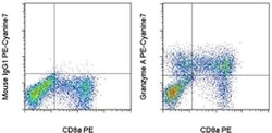
- Normal human peripheral blood cells were stimulated for 3 days with Human IL-2 Recombinant Protein (Product # 14-8029-81), then cultured with Protein Transport Inhibitor Cocktail (Product # 00-4980-03) for an additional 5 hours. The cells were intracellularly stained with Anti-Human CD8a PE (Product # 12-0088-80) and Mouse IgG1 K Isotype Control PE-Cyanine7 (Product # 25-4714-80) (left) or Anti-Human Granzyme A PE-Cyanine7 (right) using the Intracellular Fixation and Permeabilization Buffer Set (Product # 88-8824-00) and protocol. Cells in the lymphocyte gate were used for analysis.

Supportive validation
- Submitted by
- Invitrogen Antibodies (provider)
- Main image
- Experimental details
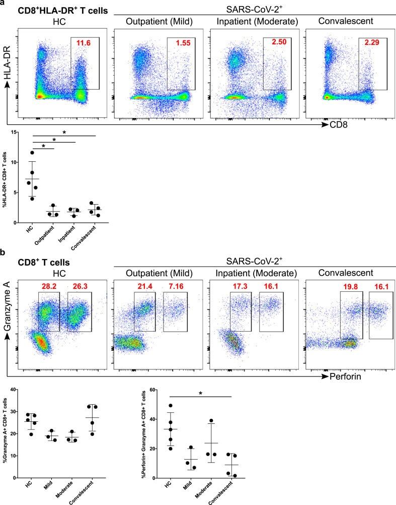
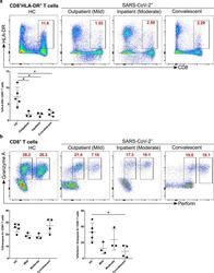
- Figure 6 Decreased activation and cytotoxic functional protein expression of CD8 + T cells in convalescent patients. A. CD8 + T cells were examined for the expression of activation marker HLA-DR (upper FACS panel). One exemplary dot plot is shown per study group. The bar diagram (lower panel) shows that HLA-DR was a significantly lower on CD8 + T cells in mild, moderate and convalescent COVID-19 + patients compared with HC. *P-value