Explore
 Validate
 Learn
Learn Flow cytometry
Antibody data
- Antibody Data
- Antigen structure
- References [3]
- Comments [0]
- Validations
- Flow cytometry [1]
- Other assay [1]
Submit
Validation data
Reference
Comment
Report error
- Product number
- 50-9177-41 - Provider product page

- Provider
- Invitrogen Antibodies
- Product name
- Granzyme A Monoclonal Antibody (CB9), eFluor™ 660, eBioscience™
- Antibody type
- Monoclonal
- Antigen
- Other
- Description
- Description: This CB9 monoclonal antibody reacts with human Granzyme A. Granzyme A is the most abundantly expressed of the granzyme serine proteases, which are proteins released from the granules of NK cells and cytotoxic T lymphocytes that induce death in target cells by cleavage of intracellular substrates. They play a critical role in immune defense against viruses, tumors, and intracellular bacteria. Granzyme A activates caspase-independent cell death pathways morphologically similar to apoptosis and characterized by mitochondrial and DNA damage. It may also play a role in inflammation, as the precursor form of IL-1 beta (pro-IL-1 beta) is among its target substrates. Granzyme A shares overlapping substrate specificity with the closely-related Granzyme K, which is believed to account for the minimal decrease in cytotoxicity of Granzyme A-deficient CTL.
- Antibody clone number
- CB9
- Concentration
- 5 µL/Test
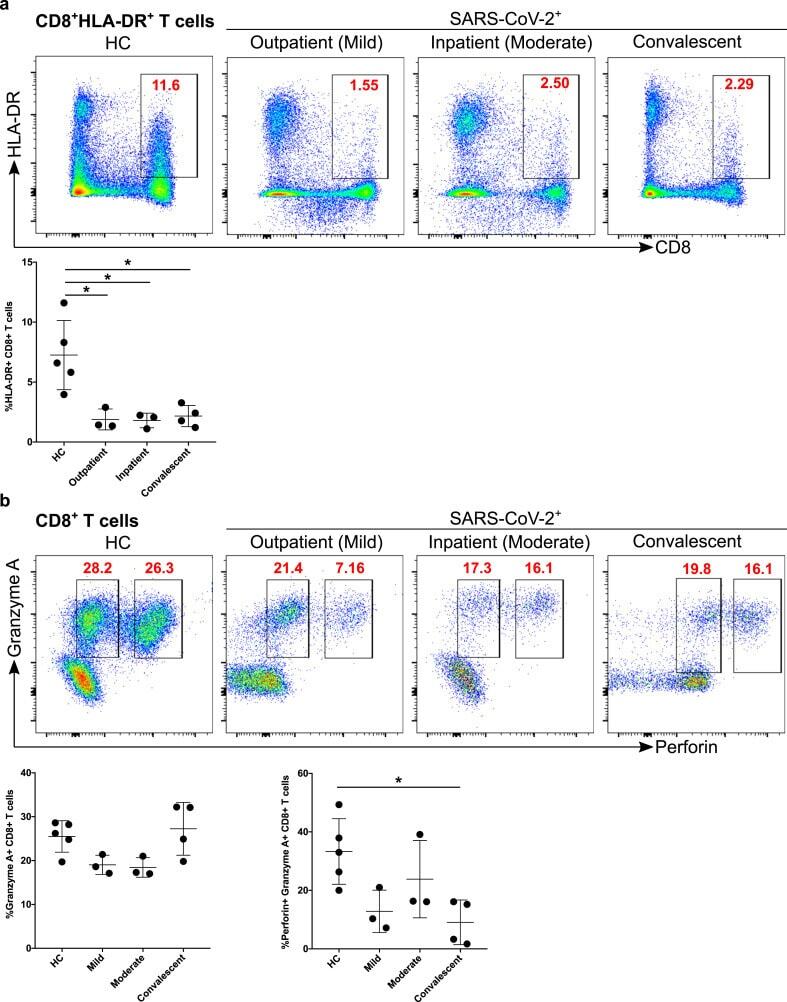
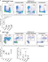
Submitted references SARS-CoV-2 infection paralyzes cytotoxic and metabolic functions of the immune cells.
Granzyme A activates another way to die.
Granzyme K displays highly restricted substrate specificity that only partially overlaps with granzyme A.
Singh Y, Trautwein C, Fendel R, Krickeberg N, Berezhnoy G, Bissinger R, Ossowski S, Salker MS, Casadei N, Riess O, Deutsche COVID-19 OMICS Initiate (DeCOI)
Heliyon 2021 Jun;7(6):e07147
Heliyon 2021 Jun;7(6):e07147
Granzyme A activates another way to die.
Lieberman J
Immunological reviews 2010 May;235(1):93-104
Immunological reviews 2010 May;235(1):93-104
Granzyme K displays highly restricted substrate specificity that only partially overlaps with granzyme A.
Bovenschen N, Quadir R, van den Berg AL, Brenkman AB, Vandenberghe I, Devreese B, Joore J, Kummer JA
The Journal of biological chemistry 2009 Feb 6;284(6):3504-12
The Journal of biological chemistry 2009 Feb 6;284(6):3504-12
No comments: Submit comment
Supportive validation
- Submitted by
- Invitrogen Antibodies (provider)
- Main image
- Experimental details
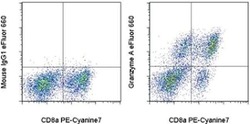
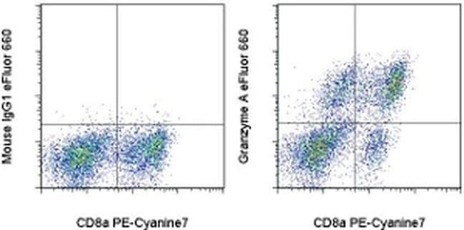
- Normal human peripheral blood cells were stimulated for 3 days with Human IL-2 Recombinant Protein (Product # 14-8029-81), then cultured with Protein Transport Inhibitor Cocktail (Product # 00-4980-03) for an additional 5 hours. The cells were stained with Anti-Human CD8a PE-Cyanine7 (Product # 25-0088-42) and Mouse IgG1 K Isotype Control eFluor® 660 (Product # 50-4714-82) (left) or Anti-Human Granzyme A eFluor® 660 (right) using the Intracellular Fixation and Permeabilization Buffer Set (Product # 88-8824-00) and protocol. Cells in the lymphocyte gate were used for analysis.
Supportive validation
- Submitted by
- Invitrogen Antibodies (provider)
- Main image
- Experimental details
- Figure 6 Decreased activation and cytotoxic functional protein expression of CD8 + T cells in convalescent patients. A. CD8 + T cells were examined for the expression of activation marker HLA-DR (upper FACS panel). One exemplary dot plot is shown per study group. The bar diagram (lower panel) shows that HLA-DR was a significantly lower on CD8 + T cells in mild, moderate and convalescent COVID-19 + patients compared with HC. *P-value