Explore
 Validate
 Learn
LearnMA1-90928
antibody from Invitrogen Antibodies
Targeting: CSPG4
CSPG4A, HMW-MAA, MCSP, MCSPG, MEL-CSPG, MSK16, NG2
 Western blot
 Immunocytochemistry
 Immunoprecipitation
Antibody data
- Antibody Data
- Antigen structure
- References [1]
- Comments [0]
- Validations
- Immunocytochemistry [1]
Submit
Validation data
Reference
Comment
Report error
- Product number
- MA1-90928 - Provider product page

- Provider
- Invitrogen Antibodies
- Product name
- NG2 Monoclonal Antibody (LHM 2)
- Antibody type
- Monoclonal
- Antigen
- Other
- Description
- MA1-90928 detects MCSP from human samples.
- Reactivity
- Human
- Host
- Mouse
- Isotype
- IgG
- Antibody clone number
- LHM 2
- Vial size
- 100 µg
- Concentration
- 1 mg/mL
- Storage
- Store at 4°C short term. For long term storage, store at -20°C, avoiding freeze/thaw cycles.
Submitted references Monitoring the systemic human memory B cell compartment of melanoma patients for anti-tumor IgG antibodies.
Gilbert AE, Karagiannis P, Dodev T, Koers A, Lacy K, Josephs DH, Takhar P, Geh JL, Healy C, Harries M, Acland KM, Rudman SM, Beavil RL, Blower PJ, Beavil AJ, Gould HJ, Spicer J, Nestle FO, Karagiannis SN
PloS one 2011 Apr 29;6(4):e19330
PloS one 2011 Apr 29;6(4):e19330
No comments: Submit comment
Supportive validation
- Submitted by
- Invitrogen Antibodies (provider)
- Main image

- Experimental details
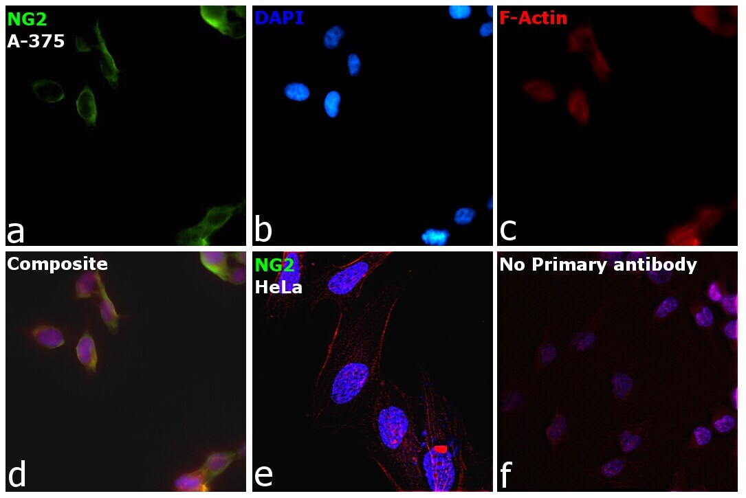
- Immunofluorescence analysis of NG2 was performed using 70% confluent log phase A-375 cells. The cells were fixed with 4% Paraformaldehyde for 10 Minutes, permeabilized with 0.1% Triton™ X-100 for 10 Minutes, and blocked with 2% BSA for 10 Minutes at room temperature. The cells were labeled with NG2 Monoclonal Antibody (LHM 2) (Product # MA1-90928) at 5 µg/mL in 0.1% BSA, incubated at 4 degree Celsius overnight and then labeled with Goat anti-Mouse IgG (H+L) Superclonal™ Recombinant Secondary Antibody, Alexa Fluor® 488 conjugate (Product # A28175, 1:2,000 dilution) for 45 minutes at room temperature (Panel a: Green). Nuclei (Panel b: Blue) were stained with SlowFade® Gold Antifade Mountant with DAPI (Product # S36938). F-actin (Panel c: Red) was stained with Rhodamine Phalloidin (Product # R415, 1:300). Panel d represents the merged image showing membrane localization. Panel e represents HeLa cells with no expression for NG2. Panel f represents control cells with no primary antibody to assess background. The images were captured at 60X magnification.