 Explore
 Validate
 Learn
LearnMAB6689
antibody from Novus Biologicals
Targeting: CSPG4
CSPG4A, HMW-MAA, MCSP, MCSPG, MEL-CSPG, MSK16, NG2
 Immunohistochemistry
Antibody data
- Antibody Data
- Antigen structure
- References [2]
- Comments [0]
- Validations
- Immunohistochemistry [1]
Submit
Validation data
Reference
Comment
Report error
- Product number
- MAB6689 - Provider product page

- Provider
- Novus Biologicals
- Product name
- Rat Monoclonal NG2/MCSP Antibody
- Antibody type
- Monoclonal
- Description
- Protein A or G purified from hybridoma culture supernatant. Detects mouse NG2/MCSP in direct ELISAs. In direct ELISAs, no cross-reactivity with recombinant human NG2/MCSP/CSPG4 is observed.
- Reactivity
- Mouse
- Host
- Rat
- Isotype
- IgG
- Vial size
- 100 ug
- Concentration
- LYOPH
- Storage
- Use a manual defrost freezer and avoid repeated freeze-thaw cycles. 12 months from date of receipt, -20 to -70 degreesC as supplied. 1 month, 2 to 8 degreesC under sterile conditions after reconstitution. 6 months, -20 to -70 degreesC under sterile conditions after reconstitution.
Submitted references Dysregulated mesenchymal PDGFR-β drives kidney fibrosis.
Silencing of galectin-1 inhibits retinal neovascularization and ameliorates retinal hypoxia in a murine model of oxygen-induced ischemic retinopathy.
Buhl EM, Djudjaj S, Klinkhammer BM, Ermert K, Puelles VG, Lindenmeyer MT, Cohen CD, He C, Borkham-Kamphorst E, Weiskirchen R, Denecke B, Trairatphisan P, Saez-Rodriguez J, Huber TB, Olson LE, Floege J, Boor P
EMBO molecular medicine 2020 Mar 6;12(3):e11021
EMBO molecular medicine 2020 Mar 6;12(3):e11021
Silencing of galectin-1 inhibits retinal neovascularization and ameliorates retinal hypoxia in a murine model of oxygen-induced ischemic retinopathy.
Yang N, Zhang W, He T, Xing Y
Experimental eye research 2017 Jun;159:1-15
Experimental eye research 2017 Jun;159:1-15
No comments: Submit comment
Supportive validation
- Submitted by
- Novus Biologicals (provider)
- Main image
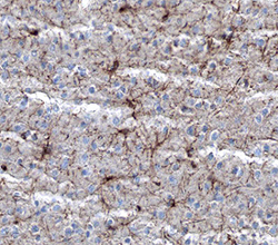
- Experimental details
- NG2/MCSP in Mouse Brain. NG2/MCSP was detected in perfusion fixed frozen sections of mouse brain (cortex) using Rat Anti-Mouse NG2/MCSP Monoclonal Antibody (Catalog # MAB6689) at 25 µg/mL overnight at 4 °C. Tissue was stained using the Anti-Rat HRP-DAB Cell & Tissue Staining Kit (brown; Catalog # CTS017) and counterstained with hemotoxylin (blue). Specific staining was localized to glial cells. View our protocol for Chromogenic IHC Staining of Frozen Tissue Sections.
