 Explore
 Validate
 Learn
Learn14-6504-82
antibody from Invitrogen Antibodies
Targeting: CSPG4
CSPG4A, HMW-MAA, MCSP, MCSPG, MEL-CSPG, MSK16, NG2
 Western blot
 Immunocytochemistry
 Immunoprecipitation Immunohistochemistry Flow cytometry Other assay
Antibody data
- Antibody Data
- Antigen structure
- References [8]
- Comments [0]
- Validations
- Immunocytochemistry [4]
- Other assay [6]
Submit
Validation data
Reference
Comment
Report error
- Product number
- 14-6504-82 - Provider product page

- Provider
- Invitrogen Antibodies
- Product name
- Neural/Glial Antigen 2 (NG2) Monoclonal Antibody (9.2.27), eBioscience™
- Antibody type
- Monoclonal
- Antigen
- Other
- Description
- Description: This 9.2.27 monoclonal antibody reacts with human neural/glial antigen 2, which is also known as melanoma chondroitin sulfate proteoglycan (MCSP). This antigen is composed of a 250 kDa N-linked glycoprotein and a >450 kDa proteoglycan. NG2 is present on the surface of >90% of malignant melanomas in addition to some non-melanomic tumors. Moreover, NG2 is expressed on glioma cells, as well as on developing and adult oligodendrocyte precursor cells. Studies have demonstrated the involvement of this protein in tumor cell proliferation, adhesion, migration, and invasion. NG2 also functions as a coreceptor for alpha4beta1 integrin. Finally, expression of NG2 can be used as a prognostic marker for disorders such as acral letiginous melanoma and infantile acute myeloid leukemia. The 9.2.27 antibody has been reported to suppress melanoma tumor growth. Applications Reported: This 9.2.27 has been reported for use in flow cytometric analysis, immunoprecipitation, immunohistochemical staining of formalin-fixed paraffin embedded tissue (IHC-P), and immunocytochemical staining of fixed cells (ICC). Applications Tested: This 9.2.27 antibody has been tested by immunocytochemistry on methanol-fixed A375 cells at less than or equal to 10 µg/mL and by flow cytometric analysis on A375 cells at less than or equal to 0.125 µg/test. A test is defined as the amount (ug) of antibody that will stain a cell sample in a final volume of 100 µL. Cell number should be determined empirically but can range from 10e5 to 10e8 cells/test. It is recommended that this antibody be carefully titrated for optimal performance in the assay of interest. Purity: Greater than 90%, as determined by SDS-PAGE. Aggregation: Less than 10%, as determined by HPLC. Filtration: 0.2 µm post-manufacturing filtered.
- Reactivity
- Human
- Host
- Mouse
- Isotype
- IgG
- Antibody clone number
- 9.2.27
- Vial size
- 100 μg
- Concentration
- 0.5 mg/mL
- Storage
- 4°C
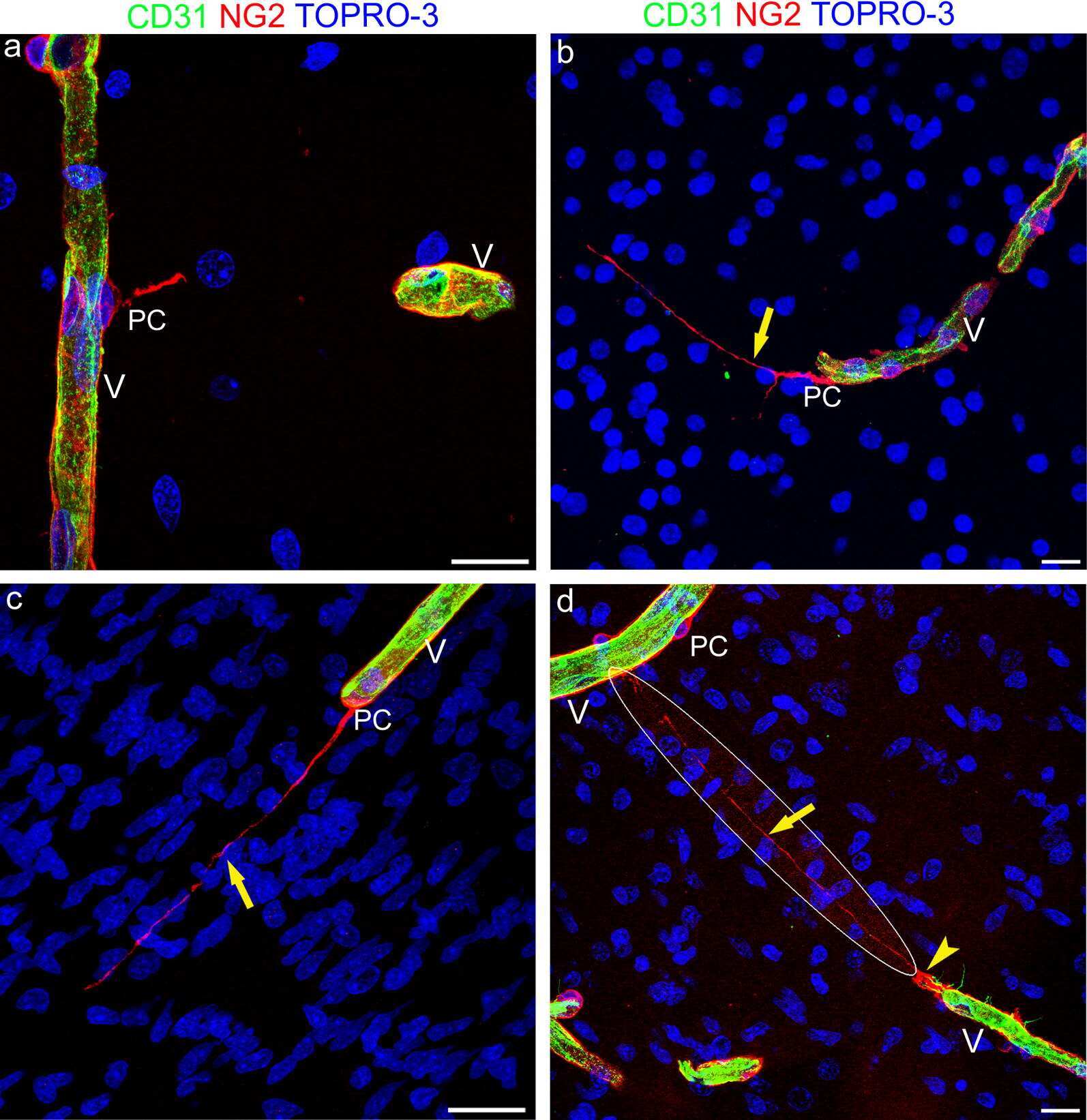
Submitted references Tunneling nanotubes evoke pericyte/endothelial communication during normal and tumoral angiogenesis.
In Vitro Modeling of Blood-Brain Barrier with Human iPSC-Derived Endothelial Cells, Pericytes, Neurons, and Astrocytes via Notch Signaling.
Melanoma chondroitin sulfate proteoglycan enhances FAK and ERK activation by distinct mechanisms.
Human uveal melanoma expresses NG2 immunoreactivity.
Molecular cloning of a human melanoma-associated chondroitin sulfate proteoglycan.
Monoclonal antibody-directed effector cells selectively lyse human melanoma cells in vitro and in vivo.
Unique glycoprotein-proteoglycan complex defined by monoclonal antibody on human melanoma cells.
Production and characterization of monoclonal antibody to a melanoma specific glycoprotein.
Errede M, Mangieri D, Longo G, Girolamo F, de Trizio I, Vimercati A, Serio G, Frei K, Perris R, Virgintino D
Fluids and barriers of the CNS 2018 Oct 5;15(1):28
Fluids and barriers of the CNS 2018 Oct 5;15(1):28
In Vitro Modeling of Blood-Brain Barrier with Human iPSC-Derived Endothelial Cells, Pericytes, Neurons, and Astrocytes via Notch Signaling.
Yamamizu K, Iwasaki M, Takakubo H, Sakamoto T, Ikuno T, Miyoshi M, Kondo T, Nakao Y, Nakagawa M, Inoue H, Yamashita JK
Stem cell reports 2017 Mar 14;8(3):634-647
Stem cell reports 2017 Mar 14;8(3):634-647
Melanoma chondroitin sulfate proteoglycan enhances FAK and ERK activation by distinct mechanisms.
Yang J, Price MA, Neudauer CL, Wilson C, Ferrone S, Xia H, Iida J, Simpson MA, McCarthy JB
The Journal of cell biology 2004 Jun 21;165(6):881-91
The Journal of cell biology 2004 Jun 21;165(6):881-91
Human uveal melanoma expresses NG2 immunoreactivity.
Li Y, Madigan MC, Lai K, Conway RM, Billson FA, Crouch R, Allen BJ
The British journal of ophthalmology 2003 May;87(5):629-32
The British journal of ophthalmology 2003 May;87(5):629-32
Molecular cloning of a human melanoma-associated chondroitin sulfate proteoglycan.
Pluschke G, Vanek M, Evans A, Dittmar T, Schmid P, Itin P, Filardo EJ, Reisfeld RA
Proceedings of the National Academy of Sciences of the United States of America 1996 Sep 3;93(18):9710-5
Proceedings of the National Academy of Sciences of the United States of America 1996 Sep 3;93(18):9710-5
Monoclonal antibody-directed effector cells selectively lyse human melanoma cells in vitro and in vivo.
Schulz G, Bumol TF, Reisfeld RA
Proceedings of the National Academy of Sciences of the United States of America 1983 Sep;80(17):5407-11
Proceedings of the National Academy of Sciences of the United States of America 1983 Sep;80(17):5407-11
Unique glycoprotein-proteoglycan complex defined by monoclonal antibody on human melanoma cells.
Bumol TF, Reisfeld RA
Proceedings of the National Academy of Sciences of the United States of America 1982 Feb;79(4):1245-9
Proceedings of the National Academy of Sciences of the United States of America 1982 Feb;79(4):1245-9
Production and characterization of monoclonal antibody to a melanoma specific glycoprotein.
Morgan AC Jr, Galloway DR, Reisfeld RA
Hybridoma 1981;1(1):27-36
Hybridoma 1981;1(1):27-36
No comments: Submit comment
Supportive validation
- Submitted by
- Invitrogen Antibodies (provider)
- Main image
- Experimental details
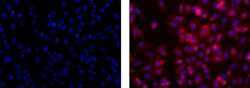
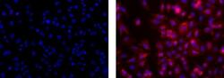
- Immunocytochemistry on fixed A375 cells using 10 µg/mL of Mouse IgG2a K Isotype control (left) or 10 µg/mL Anti-Human Neural/Glial Antigen 2 (NG2) Purified (right) followed by Anti-Mouse TRITC.Nuclei are counterstained with DAPI.
- Submitted by
- Invitrogen Antibodies (provider)
- Main image
- Experimental details
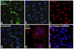
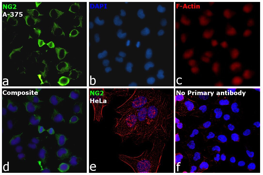
- Immunofluorescence analysis of NG2 was performed using 70% confluent log phase A-375 cells. The cells were fixed with 4% Paraformaldehyde for 10 minutes, permeabilized with 0.1% Triton™ X-100 for 10 minutes, and blocked with 2% BSA for 10 minutes at room temperature. The cells were labeled with Anti-NG2 Monoclonal Antibody (Product # 14-6504-80) at 10 µg/mL in 0.1% BSA, incubated at 4 degree Celsius overnight and then labeled with Goat anti-Mouse IgG (H+L) Superclonal™ Recombinant Secondary Antibody, Alexa Fluor® 488 conjugate (Product # A28175, 1:2000 dilution) for 45 minutes at room temperature (Panel a: Green). Nuclei (Panel b: Blue) were stained with SlowFade® Gold Antifade Mountant with DAPI (Product # S36938). F-actin (Panel c: Red) was stained with Rhodamine Phalloidin (Product # R415, 1:300). Panel d represents the merged image showing membrane localization. Panel e represents HeLa cells with no expression for NG2. Panel f represents control cells with no primary antibody to assess background. The images were captured at 60X magnification.
- Submitted by
- Invitrogen Antibodies (provider)
- Main image
- Experimental details
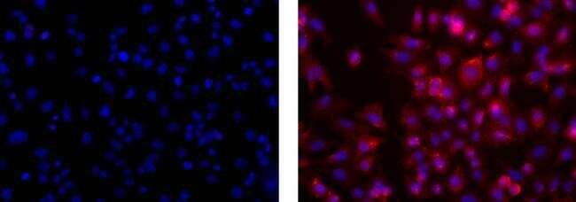
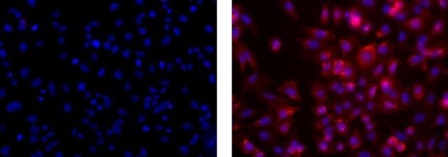
- Immunocytochemistry on fixed A375 cells using 10 µg/mL of Mouse IgG2a K Isotype control (left) or 10 µg/mL Anti-Human Neural/Glial Antigen 2 (NG2) Purified (right) followed by Anti-Mouse TRITC.Nuclei are counterstained with DAPI.
- Submitted by
- Invitrogen Antibodies (provider)
- Main image

- Experimental details
- Immunofluorescence analysis of NG2 was performed using 70% confluent log phase A-375 cells. The cells were fixed with 4% Paraformaldehyde for 10 minutes, permeabilized with 0.1% Triton™ X-100 for 10 minutes, and blocked with 2% BSA for 10 minutes at room temperature. The cells were labeled with Anti-NG2 Monoclonal Antibody (Product # 14-6504-80) at 10 µg/mL in 0.1% BSA, incubated at 4 degree Celsius overnight and then labeled with Goat anti-Mouse IgG (H+L) Superclonal™ Recombinant Secondary Antibody, Alexa Fluor® 488 conjugate (Product # A28175, 1:2000 dilution) for 45 minutes at room temperature (Panel a: Green). Nuclei (Panel b: Blue) were stained with SlowFade® Gold Antifade Mountant with DAPI (Product # S36938). F-actin (Panel c: Red) was stained with Rhodamine Phalloidin (Product # R415, 1:300). Panel d represents the merged image showing membrane localization. Panel e represents HeLa cells with no expression for NG2. Panel f represents control cells with no primary antibody to assess background. The images were captured at 60X magnification.

Supportive validation
- Submitted by
- Invitrogen Antibodies (provider)
- Main image
- Experimental details
- NULL

- Submitted by
- Invitrogen Antibodies (provider)
- Main image
- Experimental details
- NULL
- Submitted by
- Invitrogen Antibodies (provider)
- Main image
- Experimental details
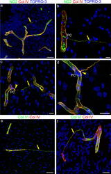
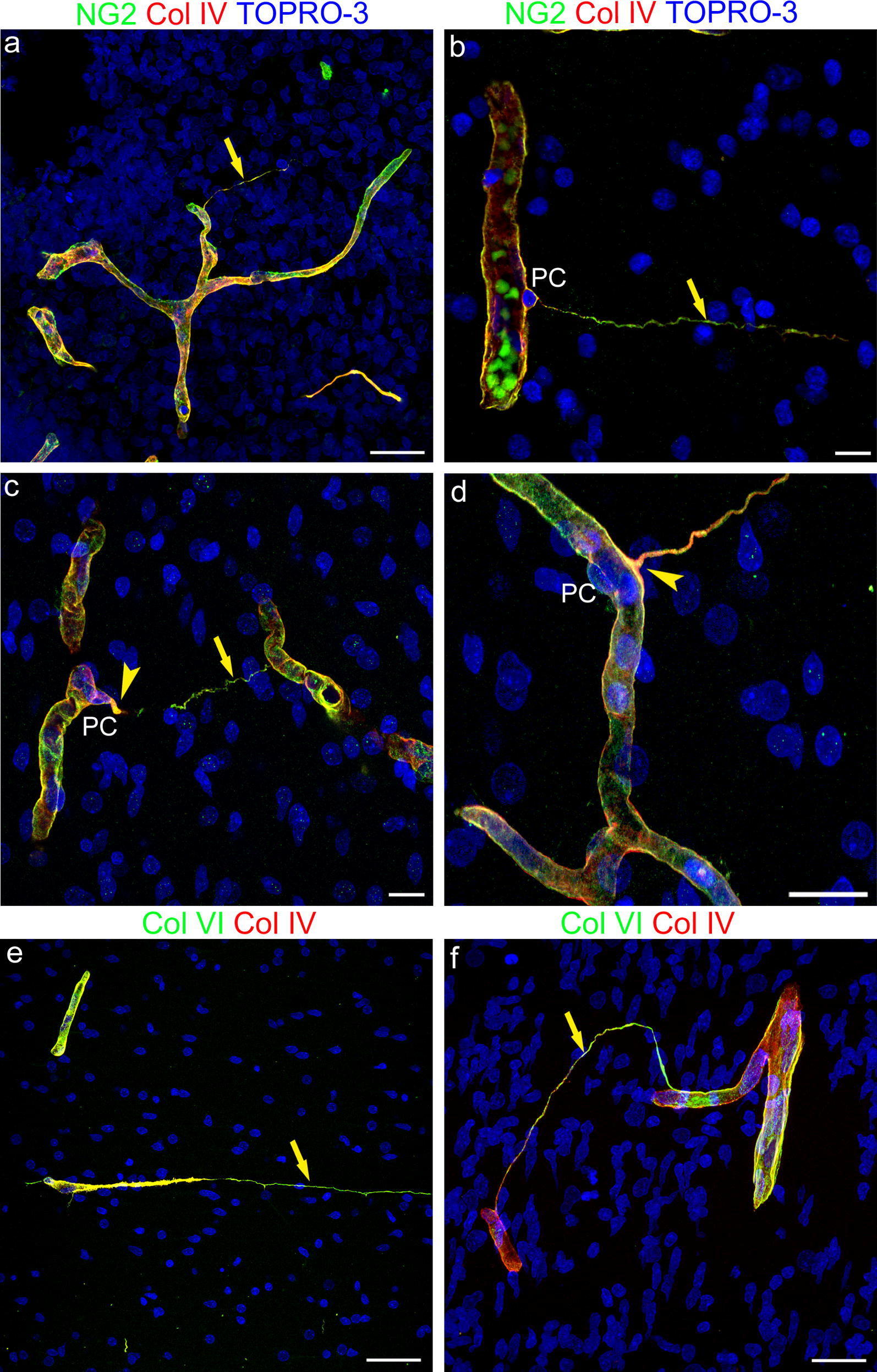
- Fig. 4 Representative confocal microscopy images of NG2/Col IV and Col VI/Col IV immunostained telencephalon sections from 18 wg ( a , f ) and 22 wg ( b - e ) fetuses. a - d NG2 and Col IV extensively colocalize on both vessel basal lamina (VBL) and TNT basal lamina (BL); NG2 staining prevails over the whole TNT extension ( a , b , c ; arrow), while Col IV appears more concentrated at the TNT root ( c , d ; arrowhead). e , f Col VI and Col IV extensively colocalize on both VBL and TNT BL, although Col VI seems to prevail on TNTs (arrow), closely resembling the NG2 staining. Nuclear counterstaining TOPRO-3. PC, pericyte. Scale bars a , d , f 25 um; b , c 20 um; e 50 um
- Submitted by
- Invitrogen Antibodies (provider)
- Main image
- Experimental details
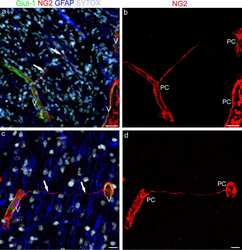
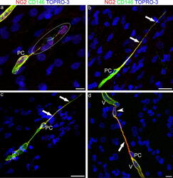
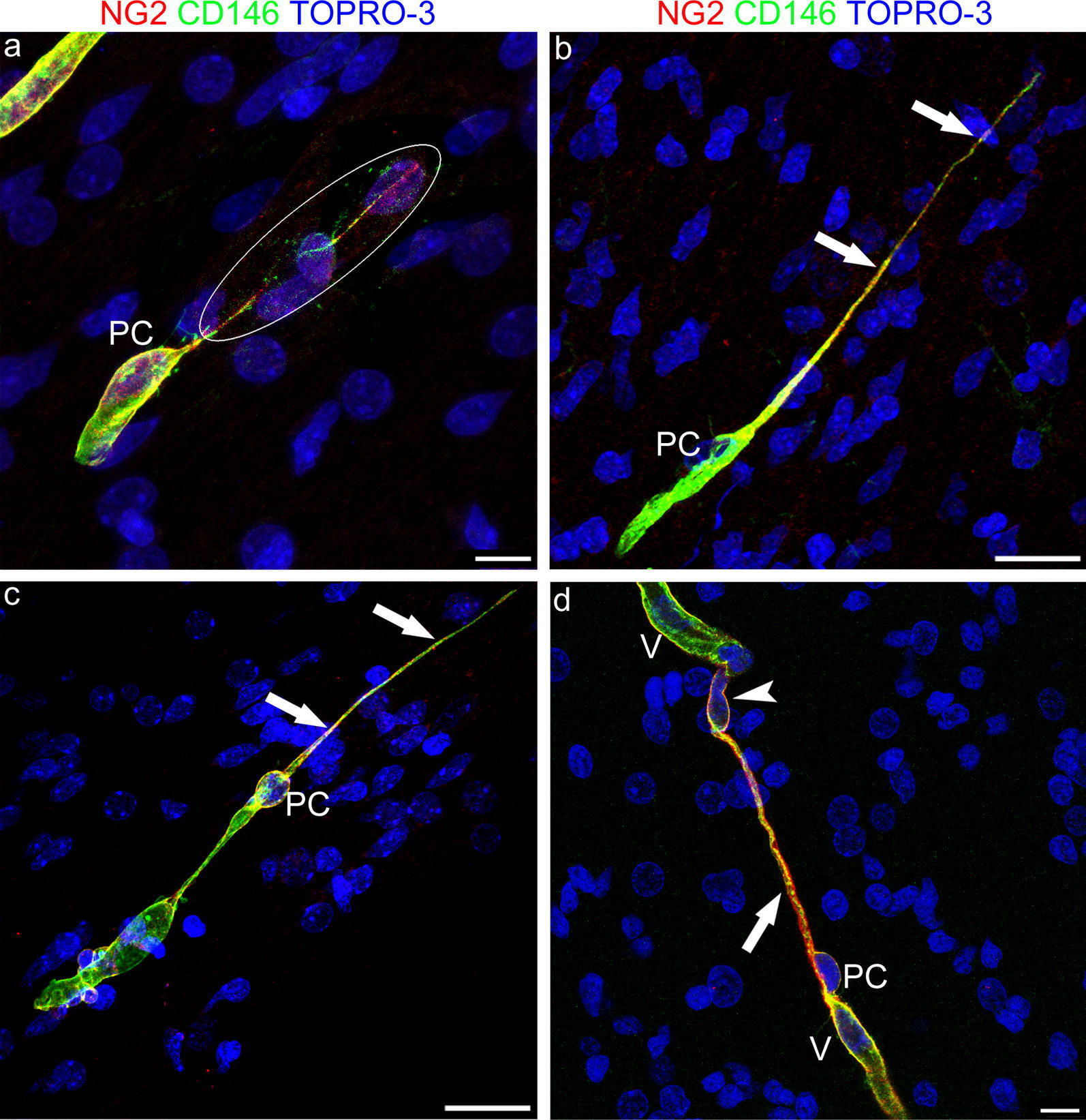
- Fig. 5 Representative confocal microscopy images of NG2/CD146 immunostained pericyte TNTs in the developing human telencephalon at 22 wg. a A very thin, double stained TNT, revealed by the overexposed laser confocal microscope setting (encircled area), originates from an NG2 + /CD146 + pericyte (PC). b , c NG2 and CD146 extensively colocalized along the entire extension of pericytes TNTs (arrows), although on the TNT distant ends CD146 staining prevails. d An NG2 + /CD146 + larger TNT conduit (arrow) appears to connect the tip of a growing microvessel (V), escorted by a leading pericyte (PC), with a recipient vessel (V); note a nucleus (arrowhead) that apparently fills the TNT end (see also Additional file 6 ). Nuclear counterstaining TOPRO-3. PC, pericyte. Scale bars a , d 10 um; b , c 25 um
- Submitted by
- Invitrogen Antibodies (provider)
- Main image
- Experimental details
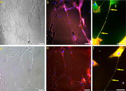
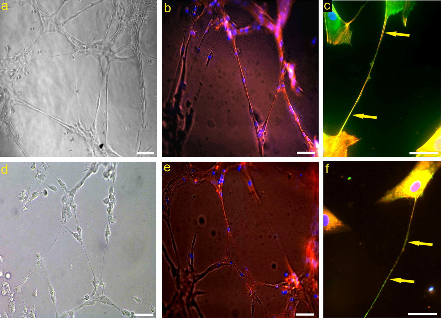
- Fig. 8 Human brain vascular pericytes (HBVP) TNTs in vitro. a , d Representative TNT networking among HBVP cultured on Matrigel layer. b , e Filaments of actin contained in HBVP-derived TNTs stained with Phalloidin TRITC-conjugated dye. c , f NG2/CSPG4 immunostaining of pericytes (green), pretreated with Phalloidin TRITC-conjugated dye ( c , red), or with lipophilic DiI cell tracker ( f , red), shows long NG2 + TNTs (arrows). Nuclear counterstaining DAPI ( b , c , e , f ). Scale bars a , b , d , e 50 um; c , f 25 um
- Submitted by
- Invitrogen Antibodies (provider)
- Main image
- Experimental details
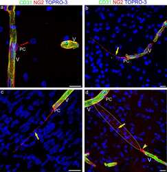
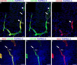
- Fig. 6 Representative confocal microscopy images of pericyte TNT involved in vessel growth and sprouting in the developing human telencephalon at 22 wg ( a - c ) and 18 wg ( d - f ). a - c CD105/CD146 and d - f NG2/CD146 double immunostainings. a - c Comparison of the merged image ( a ) with single channels ( b , c ) allows a 'complete' vessel sprout to be identified, formed by a CD146 + escorting pericyte (PC in a and b ) and a CD105 + endothelial cell (arrowhead in a , c ) and a CD146 + , pericyte-like sprout (star in a and b ), that does not stain positive for CD105, connected by a CD146 + pericyte-like TNT (arrow in a and b ); the encircled area in b corresponds to an overexposed laser confocal microscope setting. d - f NG2 colocalized with CD146 on a leading pericyte (PC) and on its short TNT (arrow). Nuclear counterstaining TOPRO-3. Scale bars a - f 25 um
