Explore
 Validate
 Learn
LearnSTJ13100192
antibody from St John's Laboratory
Targeting: CSPG4
CSPG4A, HMW-MAA, MCSP, MCSPG, MEL-CSPG, MSK16, NG2
 Western blot
 Immunohistochemistry
Antibody data
- Antibody Data
- Antigen structure
- References [0]
- Comments [0]
- Validations
- Immunohistochemistry [20]
Submit
Validation data
Reference
Comment
Report error
- Product number
- STJ13100192 - Provider product page

- Provider
- St John's Laboratory
- Product name
- Anti-NG2 antibody (ECD) (STJ13100192)
- Antibody type
- Polyclonal
- Description
- Nz White Rabbit polyclonal antibody anti-NG2 (ECD) is suitable for use in Immunohistochemistry and Western Blot research applications.
- Reactivity
- Mouse
- Conjugate
- Unconjugated
- Antigen sequence
NA- Epitope
- NA
- Isotype
- IgG
- Antibody clone number
- NA
- Vial size
- NA
- Concentration
- NA
- Storage
- Maintain the lyophilised/reconstituted antibodies frozen at-20°C for long term storage and refrigerated at 2-8°C for a shorter term. When reconstituting, glycerol (1:1) may be added for an additional stability. Avoid freeze and thaw cycles.
- Handling
- NA
No comments: Submit comment
Supportive validation
Supportive validation
Supportive validation
Supportive validation
Supportive validation
Supportive validation
Supportive validation
Supportive validation
Supportive validation
Supportive validation
Supportive validation
Supportive validation
Supportive validation
Supportive validation
Supportive validation
Supportive validation
Supportive validation
Supportive validation
Supportive validation
Supportive validation
- Submitted by
- St John's Laboratory (provider)
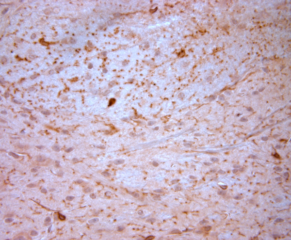
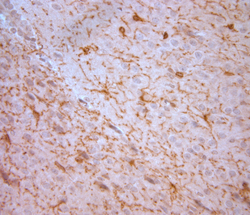
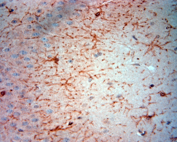
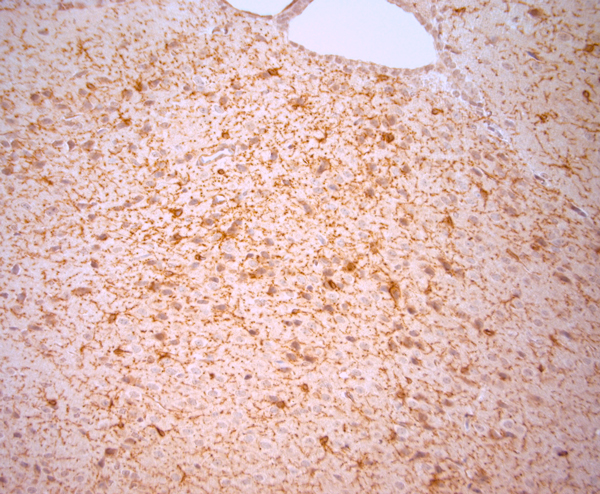
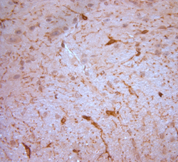
- Main image
- Experimental details
- IHC-P on paraffin sections of mouse brain. The animal was perfused using Autoperfuser at a pressure of 130 mmHg with 300 ml 4% PFA being processed for paraffin embedding. HIER: Tris-EDTA, pH 9 for 20 min using Thermo PT Module. Blocking: 0.2% LFDM in TBST filtered thru 0. 2 um. Detection was done using Novolink HRP polymer from Leica following manufacturer's instructions; DAB chromogen. Primary antibody dilution 1: 250, incubated 30 min at RT using Autostainer. Sections were counterstained with Harris Hematoxylin
- Sample type
- NA
- Validation comment
- NA
- Primary Ab dilution
- NA
- Other comments
- NA
- Secondary Ab
- NA
- Secondary Ab dilution
- NA
- Protocol
- NA
Supportive validation
- Submitted by
- St John's Laboratory (provider)
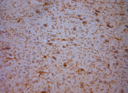
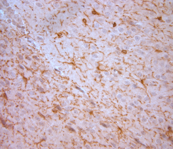
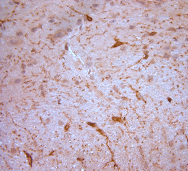
- Main image
- Experimental details
- IHC-P on paraffin sections of mouse brain. The animal was perfused using Autoperfuser at a pressure of 130 mmHg with 300 ml 4% PFA being processed for paraffin embedding. HIER: Tris-EDTA, pH 9 for 20 min using Thermo PT Module. Blocking: 0.2% LFDM in TBST filtered thru 0. 2 um. Detection was done using Novolink HRP polymer from Leica following manufacturer's instructions; DAB chromogen. Primary antibody dilution 1: 250, incubated 30 min at RT using Autostainer. Sections were counterstained with Harris Hematoxylin
- Sample type
- NA
- Validation comment
- NA
- Primary Ab dilution
- NA
- Other comments
- NA
- Secondary Ab
- NA
- Secondary Ab dilution
- NA
- Protocol
- NA

Supportive validation
- Submitted by
- St John's Laboratory (provider)
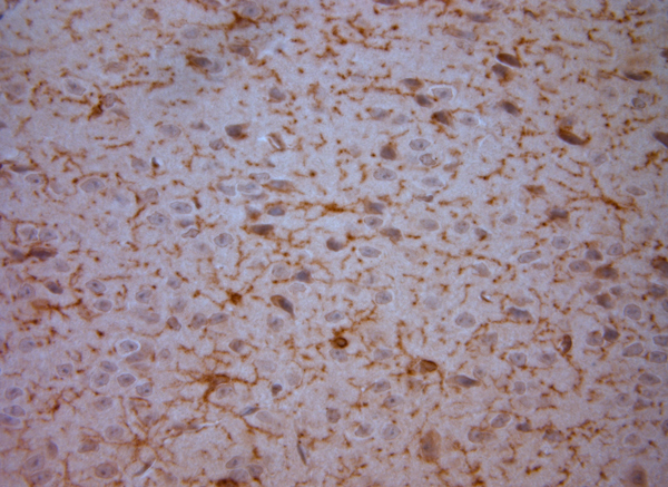
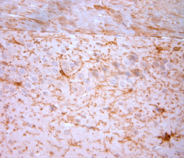
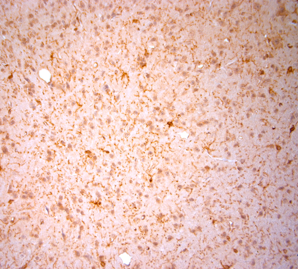
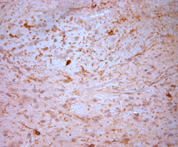
- Main image
- Experimental details
- IHC-P on paraffin sections of mouse brain. The animal was perfused using Autoperfuser at a pressure of 130 mmHg with 300 ml 4% PFA being processed for paraffin embedding. HIER: Tris-EDTA, pH 9 for 20 min using Thermo PT Module. Blocking: 0.2% LFDM in TBST filtered thru 0. 2 um. Detection was done using Novolink HRP polymer from Leica following manufacturer's instructions; DAB chromogen. Primary antibody dilution 1: 250, incubated 30 min at RT using Autostainer. Sections were counterstained with Harris Hematoxylin
- Sample type
- NA
- Validation comment
- NA
- Primary Ab dilution
- NA
- Other comments
- NA
- Secondary Ab
- NA
- Secondary Ab dilution
- NA
- Protocol
- NA
Supportive validation
- Submitted by
- St John's Laboratory (provider)
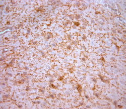
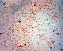
- Main image

- Experimental details
- IHC-P on paraffin sections of mouse brain. The animal was perfused using Autoperfuser at a pressure of 130 mmHg with 300 ml 4% PFA being processed for paraffin embedding. HIER: Tris-EDTA, pH 9 for 20 min using Thermo PT Module. Blocking: 0.2% LFDM in TBST filtered thru 0. 2 um. Detection was done using Novolink HRP polymer from Leica following manufacturer's instructions; DAB chromogen. Primary antibody dilution 1: 250, incubated 30 min at RT using Autostainer. Sections were counterstained with Harris Hematoxylin
- Sample type
- NA
- Validation comment
- NA
- Primary Ab dilution
- NA
- Other comments
- NA
- Secondary Ab
- NA
- Secondary Ab dilution
- NA
- Protocol
- NA
Supportive validation
- Submitted by
- St John's Laboratory (provider)
- Main image
- Experimental details
- IHC-P on paraffin sections of mouse brain. The animal was perfused using Autoperfuser at a pressure of 130 mmHg with 300 ml 4% PFA being processed for paraffin embedding. HIER: Tris-EDTA, pH 9 for 20 min using Thermo PT Module. Blocking: 0.2% LFDM in TBST filtered thru 0. 2 um. Detection was done using Novolink HRP polymer from Leica following manufacturer's instructions; DAB chromogen. Primary antibody dilution 1: 250, incubated 30 min at RT using Autostainer. Sections were counterstained with Harris Hematoxylin
- Sample type
- NA
- Validation comment
- NA
- Primary Ab dilution
- NA
- Other comments
- NA
- Secondary Ab
- NA
- Secondary Ab dilution
- NA
- Protocol
- NA
Supportive validation
- Submitted by
- St John's Laboratory (provider)
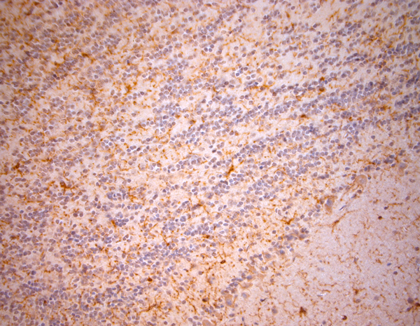
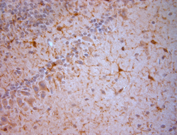
- Main image
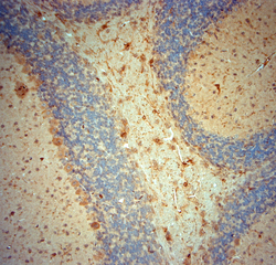
- Experimental details
- IHC-P on paraffin sections of mouse cerebellum. The animal was perfused using Autoperfuser at a pressure of 130 mmHg with 300 ml 4% PFA being processed for paraffin embedding. HIER: Tris-EDTA, pH 9 for 20 min using Thermo PT Module. Blocking: 0.2% LFDM in TBST filtered thru 0. 2 um. Detection was done using Novolink HRP polymer from Leica following manufacturer's instructions; DAB chromogen. Primary antibody dilution 1: 250, incubated 30 min at RT using Autostainer. Sections were counterstained with Harris Hematoxylin
- Sample type
- NA
- Validation comment
- NA
- Primary Ab dilution
- NA
- Other comments
- NA
- Secondary Ab
- NA
- Secondary Ab dilution
- NA
- Protocol
- NA

Supportive validation
- Submitted by
- St John's Laboratory (provider)
- Main image
- Experimental details
- IHC-P on paraffin sections of mouse cerebellum. The animal was perfused using Autoperfuser at a pressure of 130 mmHg with 300 ml 4% PFA being processed for paraffin embedding. HIER: Tris-EDTA, pH 9 for 20 min using Thermo PT Module. Blocking: 0.2% LFDM in TBST filtered thru 0. 2 um. Detection was done using Novolink HRP polymer from Leica following manufacturer's instructions; DAB chromogen. Primary antibody dilution 1: 250, incubated 30 min at RT using Autostainer. Sections were counterstained with Harris Hematoxylin
- Sample type
- NA
- Validation comment
- NA
- Primary Ab dilution
- NA
- Other comments
- NA
- Secondary Ab
- NA
- Secondary Ab dilution
- NA
- Protocol
- NA

Supportive validation
- Submitted by
- St John's Laboratory (provider)
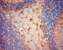
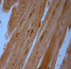
- Main image

- Experimental details
- IHC-P on paraffin sections of mouse heart. The animal was perfused using Autoperfuser at a pressure of 130 mmHg with 300 ml 4% PFA being processed for paraffin embedding. HIER: Tris-EDTA, pH 9 for 20 min using Thermo PT Module. Blocking: 0.2% LFDM in TBST filtered thru 0. 2 um. Detection was done using Novolink HRP polymer from Leica following manufacturer's instructions; DAB chromogen. Primary antibody dilution 1: 250, incubated 30 min at RT using Autostainer. Sections were counterstained with Harris Hematoxylin
- Sample type
- NA
- Validation comment
- NA
- Primary Ab dilution
- NA
- Other comments
- NA
- Secondary Ab
- NA
- Secondary Ab dilution
- NA
- Protocol
- NA
Supportive validation
- Submitted by
- St John's Laboratory (provider)
- Main image
- Experimental details
- IHC-P on paraffin sections of mouse heart. The animal was perfused using Autoperfuser at a pressure of 130 mmHg with 300 ml 4% PFA being processed for paraffin embedding. HIER: Tris-EDTA, pH 9 for 20 min using Thermo PT Module. Blocking: 0.2% LFDM in TBST filtered thru 0. 2 um. Detection was done using Novolink HRP polymer from Leica following manufacturer's instructions; DAB chromogen. Primary antibody dilution 1: 250, incubated 30 min at RT using Autostainer. Sections were counterstained with Harris Hematoxylin
- Sample type
- NA
- Validation comment
- NA
- Primary Ab dilution
- NA
- Other comments
- NA
- Secondary Ab
- NA
- Secondary Ab dilution
- NA
- Protocol
- NA

Supportive validation
- Submitted by
- St John's Laboratory (provider)
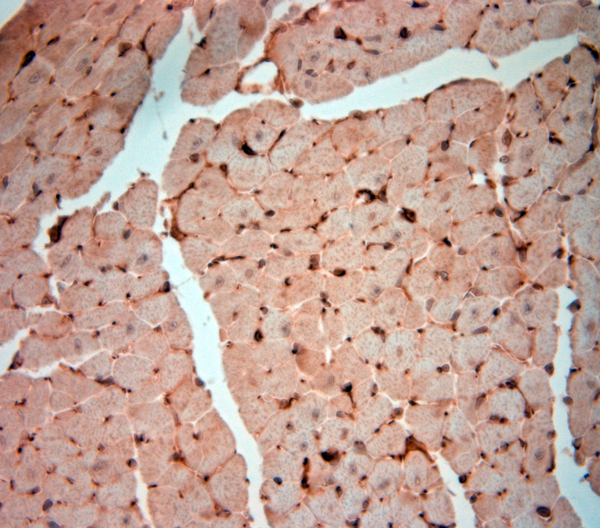
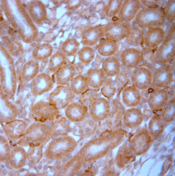
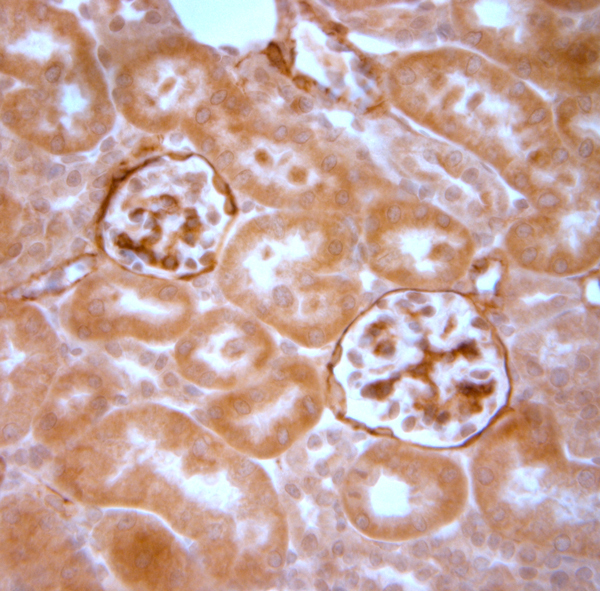
- Main image
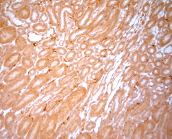
- Experimental details
- IHC-P on paraffin sections of mouse kidney. The animal was perfused using Autoperfuser at a pressure of 130 mmHg with 300 ml 4% PFA being processed for paraffin embedding. HIER: Tris-EDTA, pH 9 for 20 min using Thermo PT Module. Blocking: 0.2% LFDM in TBST filtered thru 0. 2 um. Detection was done using Novolink HRP polymer from Leica following manufacturer's instructions; DAB chromogen. Primary antibody dilution 1: 250, incubated 30 min at RT using Autostainer. Sections were counterstained with Harris Hematoxylin
- Sample type
- NA
- Validation comment
- NA
- Primary Ab dilution
- NA
- Other comments
- NA
- Secondary Ab
- NA
- Secondary Ab dilution
- NA
- Protocol
- NA

Supportive validation
- Submitted by
- St John's Laboratory (provider)
- Main image

- Experimental details
- IHC-P on paraffin sections of mouse kidney. The animal was perfused using Autoperfuser at a pressure of 130 mmHg with 300 ml 4% PFA being processed for paraffin embedding. HIER: Tris-EDTA, pH 9 for 20 min using Thermo PT Module. Blocking: 0.2% LFDM in TBST filtered thru 0. 2 um. Detection was done using Novolink HRP polymer from Leica following manufacturer's instructions; DAB chromogen. Primary antibody dilution 1: 250, incubated 30 min at RT using Autostainer. Sections were counterstained with Harris Hematoxylin
- Sample type
- NA
- Validation comment
- NA
- Primary Ab dilution
- NA
- Other comments
- NA
- Secondary Ab
- NA
- Secondary Ab dilution
- NA
- Protocol
- NA
Supportive validation
- Submitted by
- St John's Laboratory (provider)
- Main image

- Experimental details
- IHC-P on paraffin sections of mouse olfactory bulbs. The animal was perfused using Autoperfuser at a pressure of 130 mmHg with 300 ml 4% PFA being processed for paraffin embedding. HIER: Tris-EDTA, pH 9 for 20 min using Thermo PT Module. Blocking: 0.2% LFDM in TBST filtered thru 0. 2 um. Detection was done using Novolink HRP polymer from Leica following manufacturer's instructions; DAB chromogen. Primary antibody dilution 1: 250, incubated 30 min at RT using Autostainer. Sections were counterstained with Harris Hematoxylin
- Sample type
- NA
- Validation comment
- NA
- Primary Ab dilution
- NA
- Other comments
- NA
- Secondary Ab
- NA
- Secondary Ab dilution
- NA
- Protocol
- NA
Supportive validation
- Submitted by
- St John's Laboratory (provider)
- Main image
- Experimental details
- IHC-P on paraffin sections of mouse olfactory bulbs. The animal was perfused using Autoperfuser at a pressure of 130 mmHg with 300 ml 4% PFA being processed for paraffin embedding. HIER: Tris-EDTA, pH 9 for 20 min using Thermo PT Module. Blocking: 0.2% LFDM in TBST filtered thru 0. 2 um. Detection was done using Novolink HRP polymer from Leica following manufacturer's instructions; DAB chromogen. Primary antibody dilution 1: 250, incubated 30 min at RT using Autostainer. Sections were counterstained with Harris Hematoxylin
- Sample type
- NA
- Validation comment
- NA
- Primary Ab dilution
- NA
- Other comments
- NA
- Secondary Ab
- NA
- Secondary Ab dilution
- NA
- Protocol
- NA

Supportive validation
- Submitted by
- St John's Laboratory (provider)
- Main image

- Experimental details
- IHC-P on paraffin sections of mouse olfactory bulbs. The animal was perfused using Autoperfuser at a pressure of 130 mmHg with 300 ml 4% PFA being processed for paraffin embedding. HIER: Tris-EDTA, pH 9 for 20 min using Thermo PT Module. Blocking: 0.2% LFDM in TBST filtered thru 0. 2 um. Detection was done using Novolink HRP polymer from Leica following manufacturer's instructions; DAB chromogen. Primary antibody dilution 1: 250, incubated 30 min at RT using Autostainer. Sections were counterstained with Harris Hematoxylin
- Sample type
- NA
- Validation comment
- NA
- Primary Ab dilution
- NA
- Other comments
- NA
- Secondary Ab
- NA
- Secondary Ab dilution
- NA
- Protocol
- NA
Supportive validation
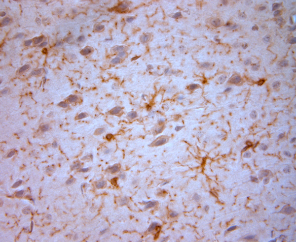
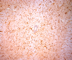
- Submitted by
- St John's Laboratory (provider)
- Main image
- Experimental details
- IHC-P on paraffin sections of mouse spinal cord. The animal was perfused using Autoperfuser at a pressure of 130 mmHg with 300 ml 4% PFA being processed for paraffin embedding. HIER: Tris-EDTA, pH 9 for 20 min using Thermo PT Module. Blocking: 0.2% LFDM in TBST filtered thru 0. 2 um. Detection was done using Novolink HRP polymer from Leica following manufacturer's instructions; DAB chromogen. Primary antibody dilution 1: 250, incubated 30 min at RT using Autostainer. Sections were counterstained with Harris Hematoxylin
- Sample type
- NA
- Validation comment
- NA
- Primary Ab dilution
- NA
- Other comments
- NA
- Secondary Ab
- NA
- Secondary Ab dilution
- NA
- Protocol
- NA

Supportive validation
- Submitted by
- St John's Laboratory (provider)
- Main image
- Experimental details
- IHC-P on paraffin sections of mouse spinal cord. The animal was perfused using Autoperfuser at a pressure of 130 mmHg with 300 ml 4% PFA being processed for paraffin embedding. HIER: Tris-EDTA, pH 9 for 20 min using Thermo PT Module. Blocking: 0.2% LFDM in TBST filtered thru 0. 2 um. Detection was done using Novolink HRP polymer from Leica following manufacturer's instructions; DAB chromogen. Primary antibody dilution 1: 250, incubated 30 min at RT using Autostainer. Sections were counterstained with Harris Hematoxylin
- Sample type
- NA
- Validation comment
- NA
- Primary Ab dilution
- NA
- Other comments
- NA
- Secondary Ab
- NA
- Secondary Ab dilution
- NA
- Protocol
- NA