Explore
 Validate
 Learn
Learn Western blot
 ELISA
ELISAAntibody data
- Antibody Data
- Antigen structure
- References [0]
- Comments [0]
- Validations
- Western blot [4]
- Immunocytochemistry [4]
- Immunohistochemistry [2]
Submit
Validation data
Reference
Comment
Report error
- Product number
- PA5-88178 - Provider product page

- Provider
- Invitrogen Antibodies
- Product name
- Aldolase C Polyclonal Antibody
- Antibody type
- Polyclonal
- Antigen
- Recombinant full-length protein
- Description
- Immunogen sequence: MPHSYPALSA EQKKELSDIA LRIVAPGKGI LAADESVGSM AKRLSQIGVE NTEENRRLYR QVLFSADDRV KKCIGGVIFF HETLYQKDDN GVPFVRTIQD KGIVVGIKVD KGVVPLAGTD GETTTQGLDG LSERCAQYKK DGADFAKWRC VLKISERTPS ALAILENANV; Positive Samples: U-87MG, LO2, Mouse brain, Mouse kidney, Rat brain, Rat spinal cord
- Reactivity
- Human, Mouse, Rat
- Host
- Rabbit
- Isotype
- IgG
- Vial size
- 100 μL
- Concentration
- 3.51 mg/mL
- Storage
- -20°C, Avoid Freeze/Thaw Cycles
No comments: Submit comment
Supportive validation
- Submitted by
- Invitrogen Antibodies (provider)
- Main image

- Experimental details
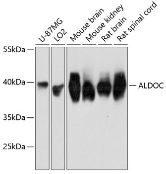
- Western blot analysis of Aldolase C in extracts of various cell lines. Samples were incubated with Aldolase C Polyclonal antibody (Product # PA5-88178) using a dilution of 1:3,000, followed by HRP Goat Anti-Rabbit IgG (H+L) at a dilution of 1:10,000. Lysates/proteins: 25 µg per lane. Blocking buffer: 3% nonfat dry milk in TBST. Detection: ECL Basic Kit. Exposure time: 3s.
- Submitted by
- Invitrogen Antibodies (provider)
- Main image
- Experimental details
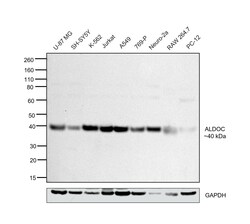
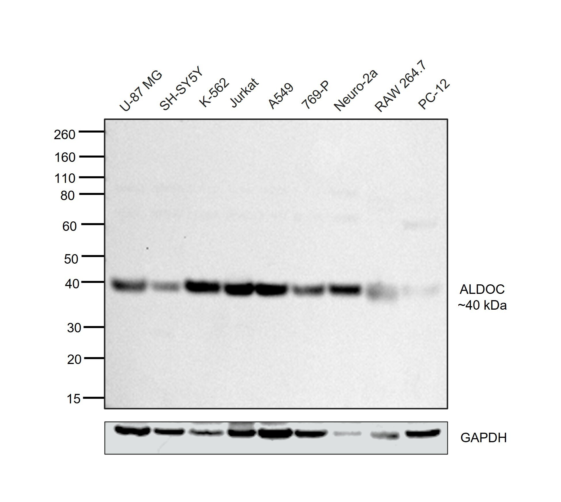
- Western blot was performed using Aldolase C Polyclonal Antibody (Product # PA5-88178) and a 40 kDa band corresponding to ALDOC was observed across cell lines tested. Whole cell extracts (30 µg lysate) of U-87 MG (Lane 1), SH-SY5Y (Lane 2), K-562 (Lane 3), Jurkat (Lane 4), A549 (Lane 5), 769-P (Lane 6), Neuro-2a (Lane 7), RAW 264.7 (Lane 8) and PC-12 (Lane 9) were electrophoresed using NuPAGE™ 4-12% Bis-Tris Protein Gel (Product # NP0321BOX), 10 well. Resolved proteins were then transferred onto a nitrocellulose membrane (Product # IB23001) by iBlot® 2 Dry Blotting System (Product # IB21001). The blot was probed with the primary antibody (1:1000) and detected by chemiluminescence with Goat anti-Rabbit IgG (H+L) Superclonal™ Recombinant Secondary Antibody, HRP (Product # A27036, 1:20,000) using the iBright™ FL1500 Imaging System (Product # A44115). Chemiluminescent detection was performed using SuperSignal™ West Pico PLUS Chemiluminescent Substrate (Product # 34580).
- Submitted by
- Invitrogen Antibodies (provider)
- Main image
- Experimental details
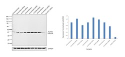
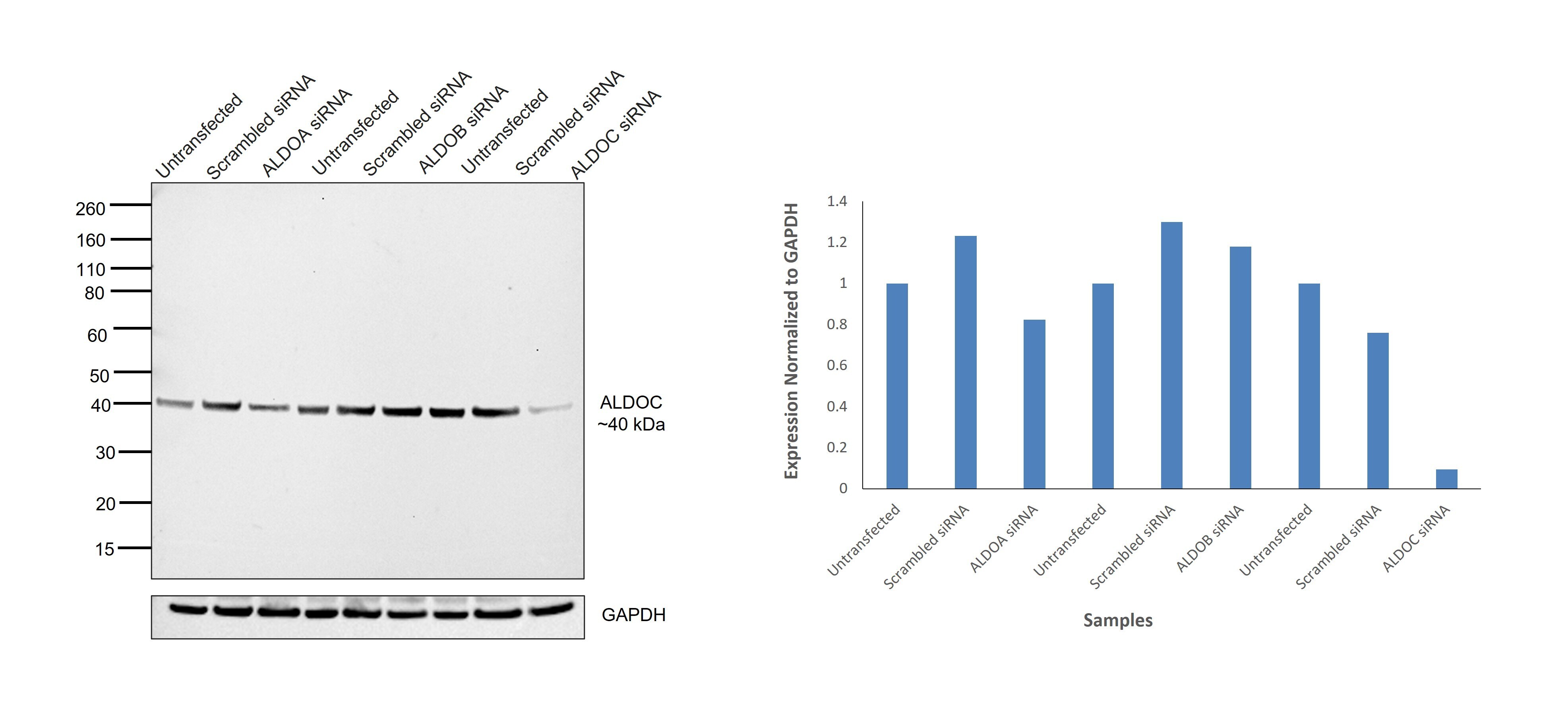
- Knockdown of ALDOC was achieved by transfecting U-87 MG with ALDOC specific siRNAs (Silencer® select Product # s1263 and s1265). Western blot analysis (Fig. a) was performed using Whole cell extracts from the ALDOC knockdown cells (lane 9), non-targeting scrambled siRNA transfected cells (lane 8) and untransfected cells (lane 7). The blot was probed with Aldolase C Polyclonal Antibody (Product # PA5-88178, 1:10,000) and Goat anti-Rabbit IgG (H+L) Superclonal™ Recombinant Secondary Antibody, HRP (Product # A27036, 1:20,000). Densitometric analysis of this western blot is shown in histogram (Fig. b). Decrease in signal upon siRNA mediated knock down confirms that antibody is specific to ALDOC. Partial knockdown was observed with ALDOA siRNAs (s1260, s1262) and no knockdown was observed with ALDOB specific siRNAs (s71 and s72) (Lanes 3 and 6).
- Submitted by
- Invitrogen Antibodies (provider)
- Main image

- Experimental details
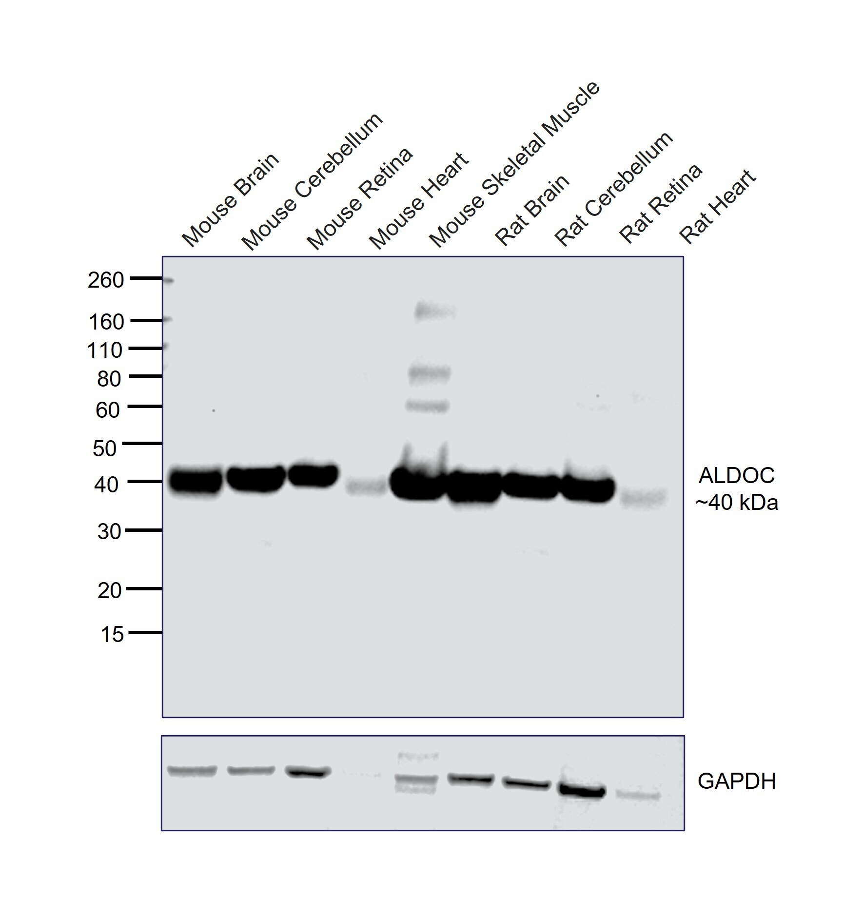
- Western blot was performed using Aldolase C Polyclonal Antibody (Product # PA5-88178) and a 40 kDa band corresponding to ALDOC was observed across tissues tested. Whole cell extracts (30 µg lysate) of Mouse Brain (Lane 1), Mouse Cerebellum (Lane 2), Mouse Retina (Lane 3), Mouse Heart (Lane 4), Mouse Skeletal Muscle (Lane 5), Rat Brain (Lane 6), Rat Cerebellum (Lane 7), Rat Retina (Lane 8) and Rat Heart (Lane 9) were electrophoresed using NuPAGE™ 4-12% Bis-Tris Protein Gel (Product # NP0321BOX), 10 well. Resolved proteins were then transferred onto a nitrocellulose membrane (Product # IB23001) by iBlot® 2 Dry Blotting System (Product # IB21001). The blot was probed with the primary antibody (1:1,000) and detected by chemiluminescence with Goat anti-Rabbit IgG (H+L) Superclonal™ Recombinant Secondary Antibody, HRP (Product # A27036, 1:20,000) using the iBright™ FL1500 Imaging System (Product # A44115). Chemiluminescent detection was performed using SuperSignal™ West Pico PLUS Chemiluminescent Substrate (Product # 34580).
Supportive validation
- Submitted by
- Invitrogen Antibodies (provider)
- Main image
- Experimental details
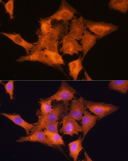
- Immunofluorescence analysis of Aldolase C in C6 cells. Samples were incubated with Aldolase C Polyclonal antibody (Product # PA5-88178) using a dilution of 1:100. Blue: DAPI for nuclear staining.
- Submitted by
- Invitrogen Antibodies (provider)
- Main image

- Experimental details
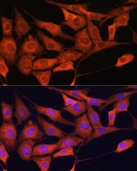
- Immunofluorescence analysis of Aldolase C in NIH/3T3 cells. Samples were incubated with Aldolase C Polyclonal antibody (Product # PA5-88178) using a dilution of 1:100. Blue: DAPI for nuclear staining.
- Submitted by
- Invitrogen Antibodies (provider)
- Main image
- Experimental details
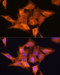
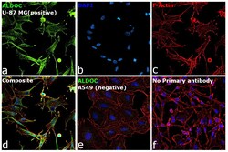
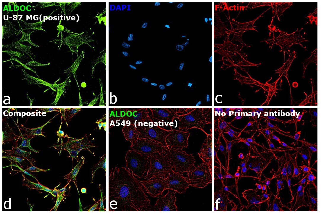
- Immunofluorescence analysis of ALDOC was performed using 70% confluent log phase U-87 MG and A549 cells. The cells were fixed with 4% paraformaldehyde for 10 minutes, permeabilized with 0.1% Triton™ X-100 for 15 minutes, and blocked with 2% BSA for 1 hour at room temperature. The cells were labeled with Aldolase C Polyclonal Antibody (Product # PA5-88178) at 1:100 in 0.1% BSA, incubated at 4 degree celsius overnight and then labeled with Donkey anti-Rabbit IgG (H+L) Highly Cross-Adsorbed Secondary Antibody, Alexa Fluor™ Plus 488 (Product # A32790), (1:2000), for 45 minutes at room temperature (Panel a: Green). Nuclei (Panel b:Blue) were stained with ProLong™ Diamond Antifade Mountant with DAPI (Product # P36962). F-actin (Panel c: Red) was stained with Rhodamine Phalloidin (Product # R415, 1:300). Panel d represents the merged image showing cytoplasmic and cytoskeletal localization in U-87 MG cells, that express ALDOC. Panel e represents the merged image showing no staining in A549 cells, that do not express ALDOC. Panel f represents control cells with no primary antibody to assess background. The images were captured at 40X magnification.
- Submitted by
- Invitrogen Antibodies (provider)
- Main image

- Experimental details
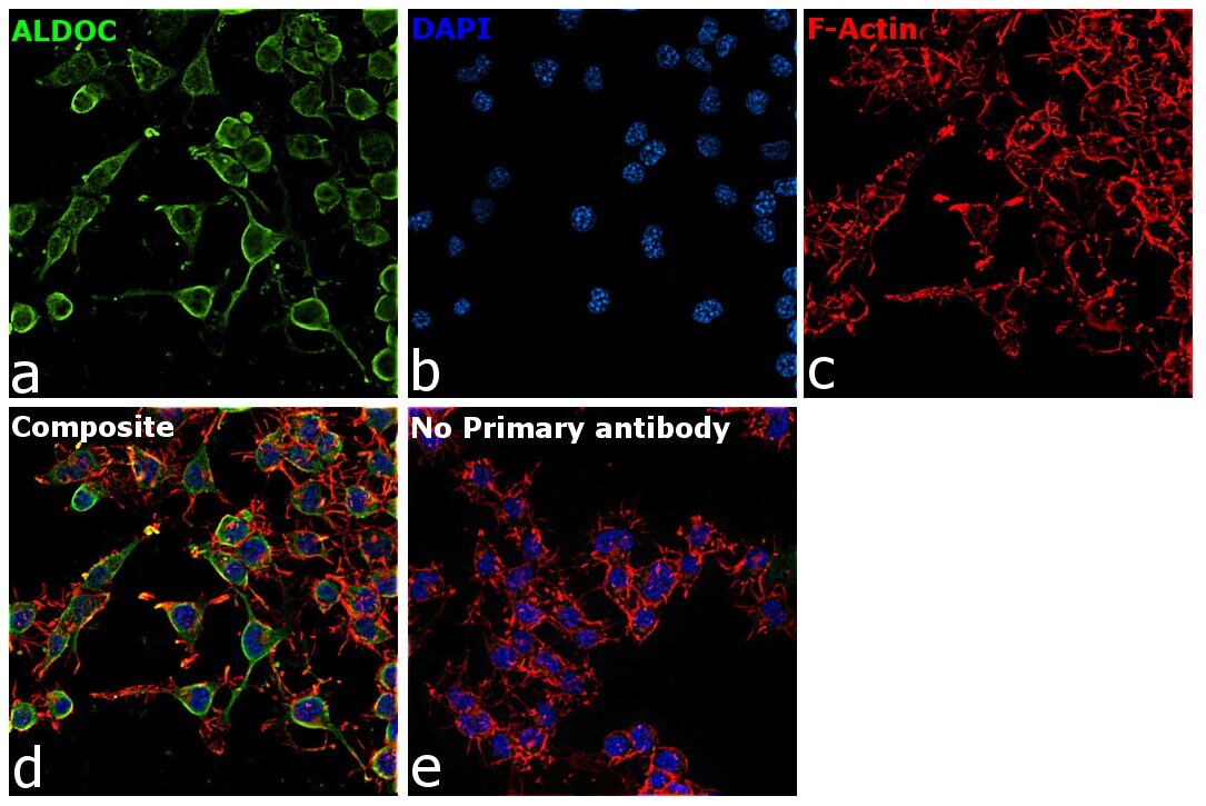
- Immunofluorescence analysis of ALDOC was performed using 70% confluent log phase Neuro-2a cells. The cells were fixed with 4% paraformaldehyde for 10 minutes, permeabilized with 0.1% Triton™ X-100 for 15 minutes, and blocked with 2% BSA for 1 hour at room temperature. The cells were labeled with Aldolase C Polyclonal Antibody (Product # PA5-88178) at 1:100 dilution in 0.1% BSA, incubated at 4 degree celsius overnight and then labeled with Donkey anti-Rabbit IgG (H+L) Highly Cross-Adsorbed Secondary Antibody, Alexa Fluor™ Plus 488 (Product # A32790), (1:2,000), for 45 minutes at room temperature (Panel a: Green). Nuclei (Panel b: Blue) were stained with ProLong™ Diamond Antifade Mountant with DAPI (Product # P36962). F-actin (Panel c: Red) was stained with Rhodamine Phalloidin (Product # R415, 1:300). Panel d represents the merged image showing membrane and cytoskeletal localization. Panel e represents control cells with no primary antibody to assess background. The images were captured at 40X magnification.
Supportive validation
- Submitted by
- Invitrogen Antibodies (provider)
- Main image
- Experimental details
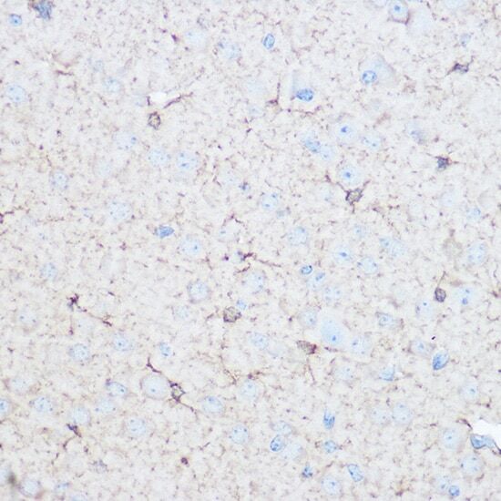
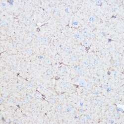
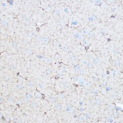
- Immunohistochemistry analysis of Aldolase C in paraffin-embedded mouse brain. Samples were incubated with Aldolase C Polyclonal antibody (Product # PA5-88178) using a dilution of 1:100 (40x lens). Perform microwave antigen retrieval with 10 mM Tris/EDTA buffer pH 9.0 before commencing with IHC staining protocol.
- Submitted by
- Invitrogen Antibodies (provider)
- Main image
- Experimental details
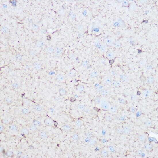
- Immunohistochemistry analysis of Aldolase C in paraffin-embedded mouse brain. Samples were incubated with Aldolase C Polyclonal antibody (Product # PA5-88178) using a dilution of 1:100 (40x lens). Perform microwave antigen retrieval with 10 mM Tris/EDTA buffer pH 9.0 before commencing with IHC staining protocol.