Explore
 Validate
 Learn
Learn Western blot
 Immunohistochemistry
Antibody data
- Antibody Data
- Antigen structure
- References [1]
- Comments [0]
- Validations
- Immunohistochemistry [1]
- Other assay [1]
Submit
Validation data
Reference
Comment
Report error
- Product number
- PA5-27659 - Provider product page

- Provider
- Invitrogen Antibodies
- Product name
- Aldolase C Polyclonal Antibody
- Antibody type
- Polyclonal
- Antigen
- Recombinant full-length protein
- Description
- Recommended positive controls: NT2D1, IMR32, U87-MG, MCF-7, mouse brain, rat brain. Predicted reactivity: Mouse (97%), Rat (97%), Xenopus laevis (83%), Dog (100%), Pig (100%), Chicken (92%), Chimpanzee (98%), Bovine (100%). Store product as a concentrated solution. Centrifuge briefly prior to opening the vial.
- Reactivity
- Human, Mouse, Rat
- Host
- Rabbit
- Isotype
- IgG
- Vial size
- 100 μL
- Concentration
- 0.28 mg/mL
- Storage
- Store at 4°C short term. For long term storage, store at -20°C, avoiding freeze/thaw cycles.
Submitted references Quantitative Proteomic Approach Reveals Altered Metabolic Pathways in Response to the Inhibition of Lysine Deacetylases in A549 Cells under Normoxia and Hypoxia.
Martín-Bernabé A, Tarragó-Celada J, Cunin V, Michelland S, Cortés R, Poignant J, Boyault C, Rachidi W, Bourgoin-Voillard S, Cascante M, Seve M
International journal of molecular sciences 2021 Mar 25;22(7)
International journal of molecular sciences 2021 Mar 25;22(7)
No comments: Submit comment
Supportive validation
- Submitted by
- Invitrogen Antibodies (provider)
- Main image
- Experimental details
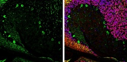
- Immunohistochemistry (Frozen) analysis of Aldolase-C was performed in frozen-sectioned adult mouse cerebellum tissue using Aldolase C Polyclonal Antibody (Product # PA5-27659) at a dilution of 1:250 (Green). Red: NeuN, stained by NeuN antibody diluted at 1:500. Blue: Fluoroshield with DAPI.

Supportive validation
- Submitted by
- Invitrogen Antibodies (provider)
- Main image
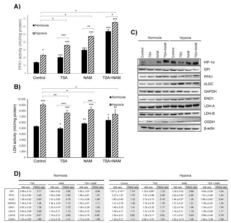
- Experimental details
- Figure 4 Effect of KDAC inhibition on enzyme activities in A549 cells under normoxia and hypoxia. ( A , B ) The ATP- dependent 6-phosphofructokinase (PFK1) ( A ) and lactate dehydrogenase (LDH) ( B ) enzymatic activities were measured after 24 h of incubation, and activities were normalized to intracellular protein content in each condition. A549 cells were treated with 1 muM of TSA, 20 mM of NAM, and both 1 muM TSA and 20 mM NAM for 24 h of incubation under normoxia and hypoxia. Cells incubated in medium without KDACIs served as control. Bars represent the means +- standard error of the mean of three independent experiments. The asterisks above bars indicate statistically significant differences compared to normoxic control cells. The asterisks above curly brackets indicate statistically significant differences between hypoxic and normoxic treatments and between hypoxic treatments and hypoxic control cells. Statistical significance was assessed by a two-tailed Student''s t -test. *, p