 Explore
 Validate
 Learn
Learn Western blot
 Immunohistochemistry
 Flow cytometry
Antibody data
- Antibody Data
- Antigen structure
- References [2]
- Comments [0]
- Validations
- Immunohistochemistry [4]
- Other assay [2]
Submit
Validation data
Reference
Comment
Report error
- Product number
- MA5-25749 - Provider product page

- Provider
- Invitrogen Antibodies
- Product name
- Heme oxygenase 2 Monoclonal Antibody (OTI1D10)
- Antibody type
- Monoclonal
- Antigen
- Recombinant full-length protein
- Reactivity
- Human
- Host
- Mouse
- Isotype
- IgG
- Antibody clone number
- OTI1D10
- Vial size
- 100 μL
- Concentration
- 0.59 mg/mL
- Storage
- -20°C, Avoid Freeze/Thaw Cycles
Submitted references Dimethyl Fumarate, an Approved Multiple Sclerosis Treatment, Reduces Brain Oxidative Stress in SIV-Infected Rhesus Macaques: Potential Therapeutic Repurposing for HIV Neuroprotection.
Regional Brain Recovery from Acute Synaptic Injury in Simian Immunodeficiency Virus-Infected Rhesus Macaques Associates with Heme Oxygenase Isoform Expression.
Garcia-Mesa Y, Xu HN, Vance P, Gruenewald AL, Garza R, Midkiff C, Alvarez-Hernandez X, Irwin DJ, Gill AJ, Kolson DL
Antioxidants (Basel, Switzerland) 2021 Mar 9;10(3)
Antioxidants (Basel, Switzerland) 2021 Mar 9;10(3)
Regional Brain Recovery from Acute Synaptic Injury in Simian Immunodeficiency Virus-Infected Rhesus Macaques Associates with Heme Oxygenase Isoform Expression.
Garcia-Mesa Y, Garza R, Diaz Ortiz ME, Gruenewald AL, Bastien BL, Lobrovich R, Irwin DJ, Betts MR, Silvestri G, Kolson DL
Journal of virology 2020 Sep 15;94(19)
Journal of virology 2020 Sep 15;94(19)
No comments: Submit comment
Supportive validation
- Submitted by
- Invitrogen Antibodies (provider)
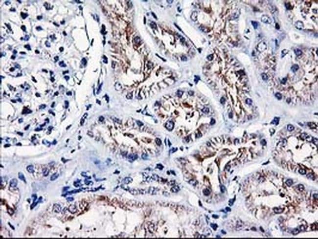
- Main image

- Experimental details
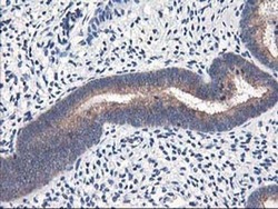
- Immunohistochemistry was performed on paraffin-embedded human kidney tissue. To expose target proteins, 10mM citric buffer, pH6.0, 100°C for 10min was used. Following antigen retrieval, tissues were probed with a HMOX2 monoclonal antibody (Product # MA5-25749).
- Submitted by
- Invitrogen Antibodies (provider)
- Main image
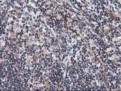
- Experimental details
- Immunohistochemistry was performed on paraffin-embedded human lymphoma tissue. To expose target proteins, 10mM citric buffer, pH6.0, 100°C for 10min was used. Following antigen retrieval, tissues were probed with a HMOX2 monoclonal antibody (Product # MA5-25749).
- Submitted by
- Invitrogen Antibodies (provider)
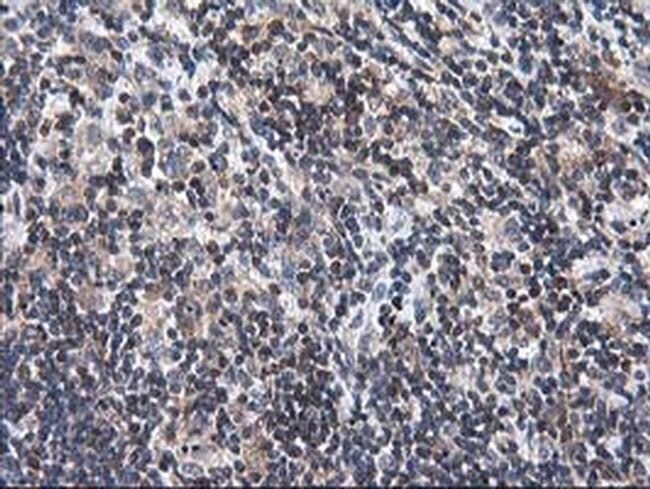
- Main image
- Experimental details
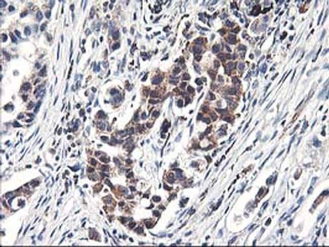
- Immunohistochemistry was performed on paraffin-embedded carcinoma of human lung tissue. To expose target proteins, 10mM citric buffer, pH6.0, 100°C for 10min was used. Following antigen retrieval, tissues were probed with a HMOX2 monoclonal antibody (Product # MA5-25749).
- Submitted by
- Invitrogen Antibodies (provider)
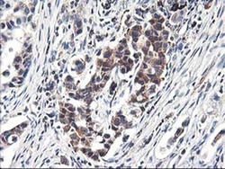
- Main image
- Experimental details
- Immunohistochemistry was performed on paraffin-embedded human endometrium tissue. To expose target proteins, 10mM citric buffer, pH6.0, 100°C for 10min was used. Following antigen retrieval, tissues were probed with a HMOX2 monoclonal antibody (Product # MA5-25749).

Supportive validation
- Submitted by
- Invitrogen Antibodies (provider)
- Main image
- Experimental details
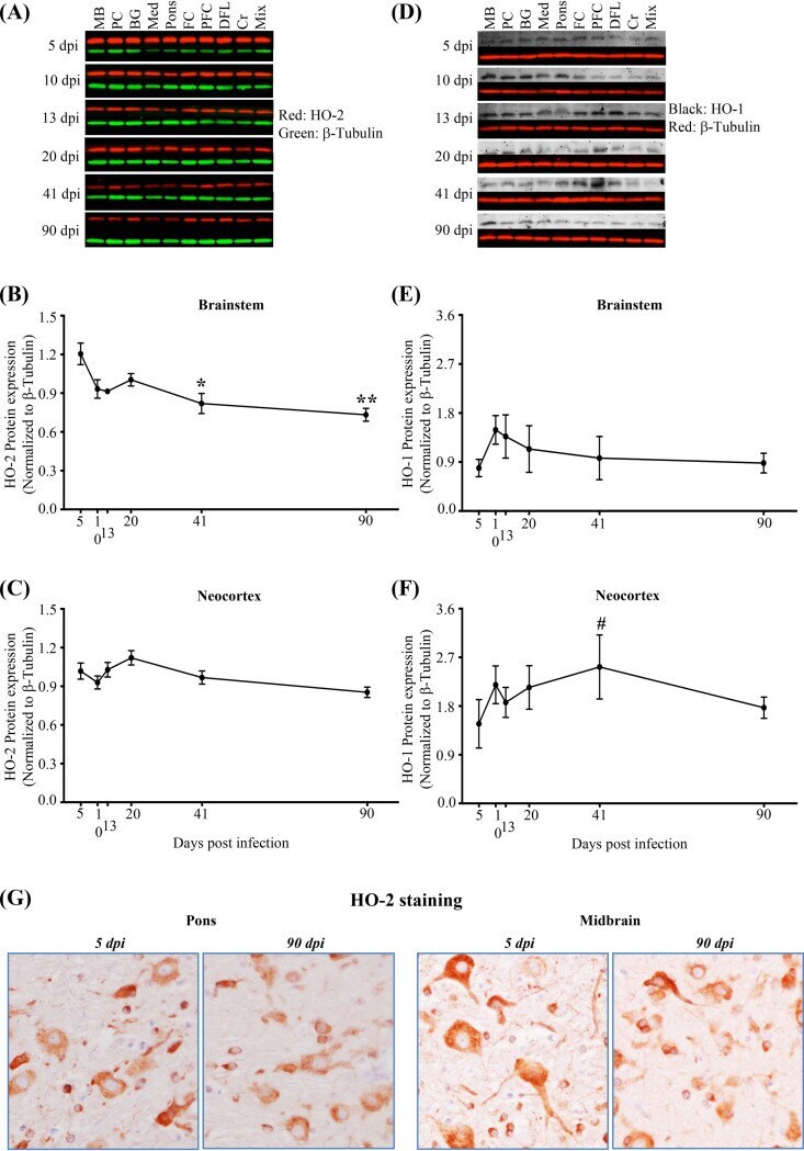
- FIG 10 SIV infection associates with progressively reduced expression of HO-2 in the brainstem and progressively increased expression of HO-1 in the neocortex. (A) Representative immunoblots of HO-2. MB, midbrain; PC, parietal cortex; BG, basal ganglia; Med, medulla; FC, frontal cortex; PFC, prefrontal cortex; DFL, deep frontal lobe; Cr, cerebellum; Mix, sample made by mixing equal volumes of all 90-dpi samples. Mix was used as a control and was run in all membranes. Each blot was normalized to that sample in each membrane. beta-Tubulin was used as a loading control. (B and C) HO-2 expression is progressively reduced in the brainstem by 41 dpi and thereafter (B), with no change in the neocortex (C). Statistical analysis was done by two-way ANOVA using repeated measures and Tukey's multiple comparisons; asterisks indicate significant differences (*, P < 0.05; **, P < 0.01) from 5 dpi. (D) Representative immunoblots of HO-1 with beta-tubulin as a loading control. (E and F) HO-1 expression is unchanged in the brainstem (E), while in the neocortex, it is increased during infection (F). Statistical analysis was done using one-way ANOVA with a test for linear trend from 5 to 41 dpi (#, P < 0.05). Each dot represents the average for 3 animals/time point (brainstem: medulla, pons, and midbrain; neocortex: parietal cortex, frontal cortex, prefrontal cortex, and deep frontal lobe). Values are means +- standard errors of the means. (G) Representative immunohistochemical staining for HO-
- Submitted by
- Invitrogen Antibodies (provider)
- Main image
- Experimental details
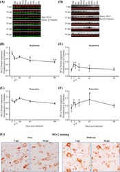
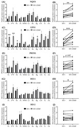
- Figure 4 DMF treatment was associated with higher antioxidant enzyme expression in the brains of SIV-infected macaques. ( A ) No significant difference in NQO1 expression was observed in the individual regions using Student's unpaired t -test (Fc: frontal cortex, 3 rd V: third ventriculi, Th: thalamus, Pc: parietal cortex, SWM: subcortical white matter, Cr: cerebellum, Bs: brainstem, Bg: basal ganglia, CN: caudate nuclei, Tc: temporal cortex, Oc: occipital cortex). ( B ) A significant increase in overall mean NQO1 expression was observed with DMF treatment (paired t -test). ( C ) A significant increase in GPX1 expression was observed in the Fc, 3 rd V, Th, Pc, and Bg with DMF treatment. ( D ) A significant increase in overall mean GPX1 expression was observed with DMF treatment. ( E ) No significant difference in PRDX1 expression was observed in individual regions. ( F ) A statistically non-significant ( p = 0.07) increase in the overall mean PRDX1 expression was observed with DMF treatment. ( G ) No significant difference in HO-1 expression was observed in individual regions. ( H ) A significant increase in overall mean HO-1 expression was observed with DMF treatment. ( I , J ) No significant difference in HO-2 expression was observed in individual regions or overall in the brains with DMF treatment. Differences in expression within individual brain regions between both groups (DMF-treated and untreated) were evaluated using Student's unpaired t -test and comparison between
