 Explore
 Validate
 Learn
Learn Western blot
 Immunohistochemistry
Antibody data
- Antibody Data
- Antigen structure
- References [1]
- Comments [0]
- Validations
- Immunohistochemistry [2]
- Other assay [1]
Submit
Validation data
Reference
Comment
Report error
- Product number
- PA5-36606 - Provider product page

- Provider
- Invitrogen Antibodies
- Product name
- RAB5C Polyclonal Antibody
- Antibody type
- Polyclonal
- Antigen
- Synthetic peptide
- Description
- Purity is >95% by SDS-PAGE.
- Reactivity
- Human, Mouse, Rat
- Host
- Rabbit
- Isotype
- IgG
- Vial size
- 100 μL
- Concentration
- 1 mg/mL
- Storage
- Store at 4°C short term. For long term storage, store at -20°C, avoiding freeze/thaw cycles.
Submitted references Paricalcitol accelerates BACE1 lysosomal degradation and inhibits calpain-1 dependent neuronal loss in APP/PS1 transgenic mice.
Fan YG, Guo T, Han XR, Liu JL, Cai YT, Xue H, Huang XS, Li YC, Wang ZY, Guo C
EBioMedicine 2019 Jul;45:393-407
EBioMedicine 2019 Jul;45:393-407
No comments: Submit comment
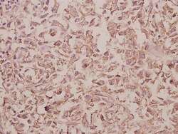
Supportive validation
- Submitted by
- Invitrogen Antibodies (provider)
- Main image
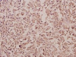
- Experimental details
- Immunohistochemical analysis of Rab 5C in paraffin-embedded human colorectal carcinoma using Rab 5C polyclonal antibody (Product # PA5-36606) at a dilution of 1:50.
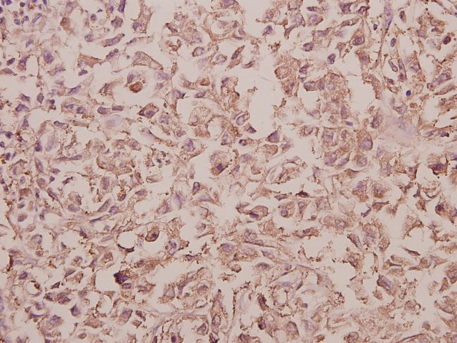
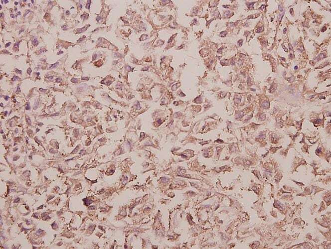
- Submitted by
- Invitrogen Antibodies (provider)
- Main image
- Experimental details
- Immunohistochemistry analysis of RAB5C in paraffin-embedded human colorectal carcinoma tissue. Samples were incubated with RAB5C polyclonal antibody (Product # PA5-36606) at a dilution of 1:50.
Supportive validation
- Submitted by
- Invitrogen Antibodies (provider)
- Main image
- Experimental details
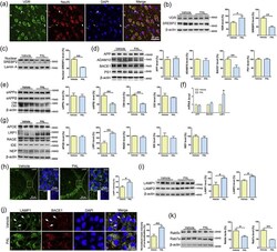
- Fig. 3 PAL treatment decreases BACE1 expression in APP/PS1 mice. (a) Sections from APP/PS1 mouse brians were co-stained with VDR (green) and NeuN (red); the merged image from cortex shows the predominant localization of VDR in NeuN-positive neurons, and the large arrows and small arrows show the localization of VDR in the epithelium and glial cells, respectively. (b-c) Inhibition of total and nuclear SREBP2 were associated with VDR activation in the cortex of APP/PS1 mice. n = 8. (d) PAL treatment dramatically suppressed the expression of BACE1 but caused no significant differences in the protein levels of APP, ADAM10 or PS1 in APP/PS1 mouse brains. n = 8. (e) Immunoblotting showed that the protein levels of sAPPbeta and C99 are decreased, but the protein levels of sAPPalpha and C83 are unchanged in APP/PS1 mouse brains after PAL treatment. n = 8. (f) APP and BACE1 mRNA expression levels were not significantly different in APP/PS1 mouse brains after PAL treatment. n = 6. (g) PAL treatment induced a marked upregulation of LRP1 without altering the expression of APOE, RAGE, IDE and NEP in APP/PS1 mouse brains. n = 8. (h) PAL treatment increased the immunointensity of LAMP1 in CA3 region. Three sections/brain, n = 6. (i) The lysosomal markers (LAMP1 and LAMP2) were increased in APP/PS1 mouse cortex after PAL treatment. n = 6. (j) Co-localizations of BACE1 and LAMP1 were increased in CA3 region after PAL treatment. The white arrows show BACE1 is not co-localized with LAMP1. Three
