 Explore
 Validate
 Learn
Learn Western blot
 Immunocytochemistry
Antibody data
- Antibody Data
- Antigen structure
- References [5]
- Comments [0]
- Validations
- Immunocytochemistry [1]
- Immunohistochemistry [1]
Submit
Validation data
Reference
Comment
Report error
- Product number
- AF1213 - Provider product page

- Provider
- R&D Systems
- Product name
- Mouse JAM-C Antibody
- Antibody type
- Polyclonal
- Description
- Immunogen affinity purified. Detects mouse JAM-C in direct ELISAs and Western blots. In Western blots, approximately 5% cross-reactivity with recombinant human JAM-C and less than 2% cross-reactivity with recombinant mouse (rm) JAM-A and rmJAM-B is observed.
- Reactivity
- Mouse
- Host
- Goat
- Conjugate
- Unconjugated
- Antigen sequence
Q9D8B7- Isotype
- IgG
- Vial size
- 100 ug
- Concentration
- LYOPH
- Storage
- Use a manual defrost freezer and avoid repeated freeze-thaw cycles. 12 months from date of receipt, -20 to -70 °C as supplied. 1 month, 2 to 8 °C under sterile conditions after reconstitution. 6 months, -20 to -70 °C under sterile conditions after reconstitution.
Submitted references Murine junctional adhesion molecules JAM-B and JAM-C mediate endothelial and stellate cell interactions during hepatic fibrosis.
Junctional adhesion molecule (JAM)-C deficient C57BL/6 mice develop a severe hydrocephalus.
Expression, localization, and function of junctional adhesion molecule-C (JAM-C) in human retinal pigment epithelium.
Junctional adhesion molecule-C promotes metastatic potential of HT1080 human fibrosarcoma.
Junctional adhesion molecules (JAM)-B and -C contribute to leukocyte extravasation to the skin and mediate cutaneous inflammation.
Hintermann E, Bayer M, Ehser J, Aurrand-Lions M, Pfeilschifter JM, Imhof BA, Christen U
Cell adhesion & migration 2016 Jul 3;10(4):419-33
Cell adhesion & migration 2016 Jul 3;10(4):419-33
Junctional adhesion molecule (JAM)-C deficient C57BL/6 mice develop a severe hydrocephalus.
Wyss L, Schäfer J, Liebner S, Mittelbronn M, Deutsch U, Enzmann G, Adams RH, Aurrand-Lions M, Plate KH, Imhof BA, Engelhardt B
PloS one 2012;7(9):e45619
PloS one 2012;7(9):e45619
Expression, localization, and function of junctional adhesion molecule-C (JAM-C) in human retinal pigment epithelium.
Economopoulou M, Hammer J, Wang F, Fariss R, Maminishkis A, Miller SS
Investigative ophthalmology & visual science 2009 Mar;50(3):1454-63
Investigative ophthalmology & visual science 2009 Mar;50(3):1454-63
Junctional adhesion molecule-C promotes metastatic potential of HT1080 human fibrosarcoma.
Fuse C, Ishida Y, Hikita T, Asai T, Oku N
The Journal of biological chemistry 2007 Mar 16;282(11):8276-83
The Journal of biological chemistry 2007 Mar 16;282(11):8276-83
Junctional adhesion molecules (JAM)-B and -C contribute to leukocyte extravasation to the skin and mediate cutaneous inflammation.
Ludwig RJ, Zollner TM, Santoso S, Hardt K, Gille J, Baatz H, Johann PS, Pfeffer J, Radeke HH, Schön MP, Kaufmann R, Boehncke WH, Podda M
The Journal of investigative dermatology 2005 Nov;125(5):969-76
The Journal of investigative dermatology 2005 Nov;125(5):969-76
No comments: Submit comment
Supportive validation
- Submitted by
- R&D Systems (provider)
- Main image
- Experimental details
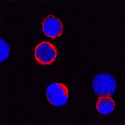
- JAM-C in Mouse Splenocytes. JAM-C was detected in immersion fixed mouse splenocytes using Goat Anti-Mouse JAM-C Antigen Affinity-purified Polyclonal Antibody (Catalog # AF1213) at 15 µg/mL for 3 hours at room temperature. Cells were stained using the NorthernLights™ 557-conjugated Anti-Goat IgG Secondary Antibody (red; Catalog # NL001) and counterstained with DAPI (blue). Specific staining was localized to cytoplasm. View our protocol for Fluorescent ICC Staining of Non-adherent Cells.

Supportive validation
- Submitted by
- R&D Systems (provider)
- Main image
- Experimental details
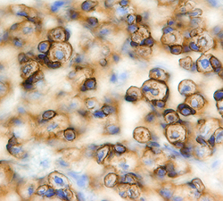
- JAM-C in Mouse Embryo. JAM-C was detected in immersion fixed frozen sections of mouse embryo (15 d.p.c.) using Goat Anti-Mouse JAM-C Antigen Affinity-purified Polyclonal Antibody (Catalog # AF1213) at 15 µg/mL overnight at 4 °C. Tissue was stained using the Anti-Goat HRP-DAB Cell & Tissue Staining Kit (brown; Catalog # CTS008) and counterstained with hematoxylin (blue). Specific staining was localized to muscle cells. View our protocol for Chromogenic IHC Staining of Frozen Tissue Sections.
