Explore
 Validate
 Learn
Learn Western blot
Antibody data
- Antibody Data
- Antigen structure
- References [3]
- Comments [0]
- Validations
- Western blot [1]
- Blocking/Neutralizing [1]
Submit
Validation data
Reference
Comment
Report error
- Product number
- MAB1189 - Provider product page

- Provider
- R&D Systems
- Product name
- Human JAM-C Antibody
- Antibody type
- Monoclonal
- Description
- Protein A or G purified from hybridoma culture supernatant. Detects human JAM-C in direct ELISAs and Western blots. In direct ELISAs and Western blots, no cross-reactivity with recombinant human JAM-A, recombinant mouse (rm) JAM-A, or rmJAM-C is observed.
- Reactivity
- Human
- Host
- Mouse
- Conjugate
- Unconjugated
- Antigen sequence
Q9BX67- Isotype
- IgG
- Antibody clone number
- 208206
- Vial size
- 500 ug
- Concentration
- LYOPH
- Storage
- Use a manual defrost freezer and avoid repeated freeze-thaw cycles. 12 months from date of receipt, -20 to -70 °C as supplied. 1 month, 2 to 8 °C under sterile conditions after reconstitution. 6 months, -20 to -70 °C under sterile conditions after reconstitution.
Submitted references Constitutive and functionally relevant expression of JAM-C on platelets.
Antibody blockade of junctional adhesion molecule-A in rabbit corneal endothelial tight junctions produces corneal swelling.
Possible involvement of gap junctions in the barrier function of tight junctions of brain and lung endothelial cells.
Erpenbeck L, Rubant S, Hardt K, Santoso S, Boehncke WH, Schön MP, Ludwig RJ
Thrombosis and haemostasis 2010 Apr;103(4):857-9
Thrombosis and haemostasis 2010 Apr;103(4):857-9
Antibody blockade of junctional adhesion molecule-A in rabbit corneal endothelial tight junctions produces corneal swelling.
Mandell KJ, Holley GP, Parkos CA, Edelhauser HF
Investigative ophthalmology & visual science 2006 Jun;47(6):2408-16
Investigative ophthalmology & visual science 2006 Jun;47(6):2408-16
Possible involvement of gap junctions in the barrier function of tight junctions of brain and lung endothelial cells.
Nagasawa K, Chiba H, Fujita H, Kojima T, Saito T, Endo T, Sawada N
Journal of cellular physiology 2006 Jul;208(1):123-32
Journal of cellular physiology 2006 Jul;208(1):123-32
No comments: Submit comment
Supportive validation
- Submitted by
- R&D Systems (provider)
- Main image

- Experimental details
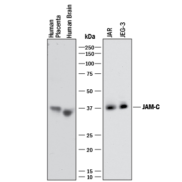
- Detection of Human JAM-C by Western Blot. Western blot shows lysates of human placenta tissue, human brain tissue, JAR human choriocarcinoma cell line, and JEG-3 human epithelial choriocarcinoma cell line. PVDF membrane was probed with 2 µg/mL of Mouse Anti-Human JAM-C Monoclonal Antibody (Catalog # MAB1189) followed by HRP-conjugated Anti-Mouse IgG Secondary Antibody (Catalog # HAF018). A specific band was detected for JAM-C at approximately 36-38 kDa (as indicated). This experiment was conducted under reducing conditions and using Immunoblot Buffer Group 1.
Supportive validation
- Submitted by
- R&D Systems (provider)
- Main image
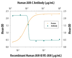
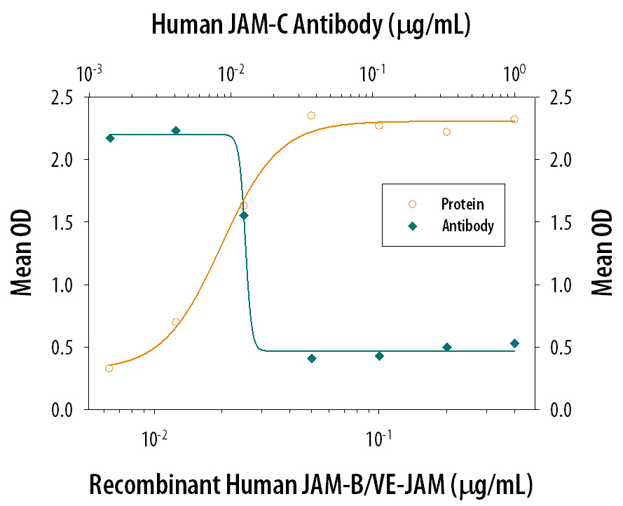
- Experimental details
- Cell Adhesion Mediated by JAM-C and Neutralization by Human JAM-C Antibody. Recombinant Human JAM-B/VE-JAM Fc Chimera, immobilized onto a microplate previously coated with Goat Anti-Human IgG Fc (Catalog # G-102-C), supports the adhesion of the J45.01 human acute lymphoblastic leukemia T lymphocyte cell line in a dose-dependent manner (orange line), as measured by endogenous cellular lysosomal acid phosphatase activity. Adhesion elicited by Recombinant Human JAM-B/VE-JAM Fc Chimera (0.2 µg/mL) is neutralized (green line) by increasing concentrations of Mouse Anti-Human JAM-C Monoclonal Antibody (Catalog # MAB1189). The ND50 is typically 0.01-0.05 µg/mL.