Explore
 Validate
 Learn
Learn Western blot
Antibody data
- Antibody Data
- Antigen structure
- References [0]
- Comments [0]
- Validations
- Western blot [2]
- Immunohistochemistry [3]
- Flow cytometry [1]
Submit
Validation data
Reference
Comment
Report error
- Product number
- ACR-042-200UL - Provider product page

- Provider
- Invitrogen Antibodies
- Product name
- CCKBR (extracellular) Polyclonal Antibody
- Antibody type
- Polyclonal
- Antigen
- Other
- Reactivity
- Human, Mouse, Rat
- Host
- Rabbit
- Isotype
- IgG
- Vial size
- 200 µL
- Concentration
- 0.8 mg/mL
- Storage
- -20° C, Avoid Freeze/Thaw Cycles
No comments: Submit comment
Supportive validation
- Submitted by
- Invitrogen Antibodies (provider)
- Main image
- Experimental details
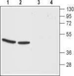
- Western blot analysis of mouse (lanes 1 and 3) and rat (lanes 2 and 4) brain membranes: - 1,2. Anti-CCKBR (extracellular) Antibody (#ACR-042), (1:200).3,4. Anti-CCKBR (extracellular) Antibody , preincubated with CCKBR (extracellular) Blocking Peptide (#BLP-CR042).

- Submitted by
- Invitrogen Antibodies (provider)
- Main image
- Experimental details
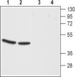
- Western blot analysis of mouse (lanes 1 and 3) and rat (lanes 2 and 4) brain membranes: - 1,2. Anti-CCKBR (extracellular) Antibody (#ACR-042), (1:200).3,4. Anti-CCKBR (extracellular) Antibody , preincubated with CCKBR (extracellular) Blocking Peptide (#BLP-CR042).
Supportive validation
- Submitted by
- Invitrogen Antibodies (provider)
- Main image
- Experimental details
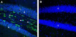
- Expression of CCKBR in mouse hippocampus. Immunohistochemical staining of perfusion-fixed frozen mouse brain sections Anti-CCKBR (extracellular) Antibody (#ACR-042), (1:200), followed by goat Anti-rabbit-AlexaFluor-488. A. Staining in the mouse hippocampal dentate gyrus region, showed immunoreactivity (green) in the granule layer (G, vertical arrows) and in interneurons (horizontal arrows). B. Pre-incubation of the Antibody with CCKBR (extracellular) Blocking Peptide (#BLP-CR042), suppressed staining. Cell nuclei are stained with DAPI (blue).
- Submitted by
- Invitrogen Antibodies (provider)
- Main image
- Experimental details
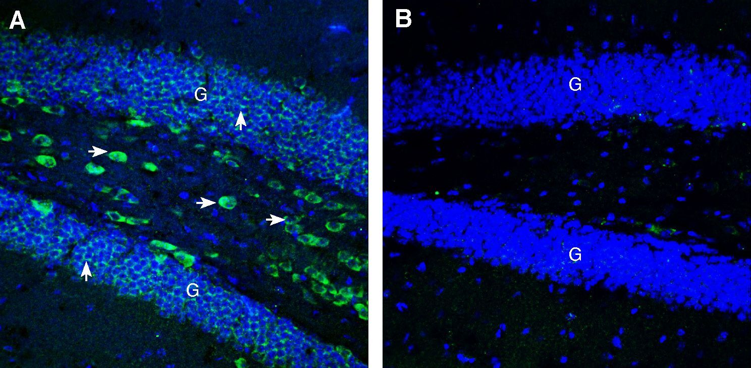
- Expression of CCKBR in rat hypothalamus. Immunohistochemical staining of perfusion-fixed frozen rat brain sections Anti-CCKBR (extracellular) Antibody (#ACR-042), (1:200), followed by goat Anti-rabbit-AlexaFluor-488. A. Staining in the rat dorsomedial hypothalamus region, showed immunoreactivity (green) in neurons (vertical arrows) and along the wall of 3rd ventricle (horizontal arrow). B. Pre-incubation of the Antibody with CCKBR (extracellular) Blocking Peptide (#BLP-CR042), suppressed staining. Cell nuclei are stained with DAPI (blue). 3rd V = Third ventricle.
- Submitted by
- Invitrogen Antibodies (provider)
- Main image
- Experimental details
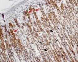
- Expression of Cholecystokinin B receptor in rat stomach - Immunohistochemical staining of paraffin embedded rat stomach sections using Anti-CCKBR (extracellular) Antibody (#ACR-042), (1:100). Cholecystokinin B receptor (brown) is expressed in both parietal cells (black arrows) and in chief cells (red arrows) of the gastric mucosa. Hematoxilin is used as the counterstain.

Supportive validation
- Submitted by
- Invitrogen Antibodies (provider)
- Main image
- Experimental details
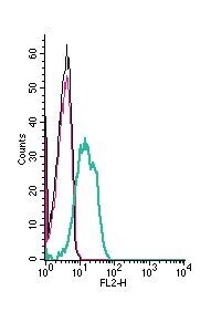
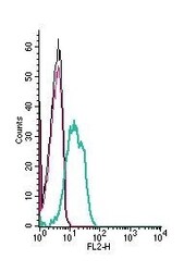
- Cell surface detection of Cholecystokinin B receptor by indirect flow cytometry in live intact human Jurkat T-cell leukemia cells: - (black line) cells. (red) Cells + goat- Anti-rabbit-PE. (green) Cells + Anti-CCKBR (extracellular) Antibody (#ACR-042), (5μg) + goat- Anti-rabbit-PE.