Explore
 Validate
 Learn
Learn Western blot
Antibody data
- Antibody Data
- Antigen structure
- References [0]
- Comments [0]
- Validations
- Western blot [1]
- Immunohistochemistry [1]
Submit
Validation data
Reference
Comment
Report error
- Product number
- PA5-18990 - Provider product page

- Provider
- Invitrogen Antibodies
- Product name
- Anti-Slc10a2
- Antibody type
- Polyclonal
- Antigen
- Synthetic peptide sequence (DETNKGFQPDEK) corresponding to the C-terminus amino acids of Slc10a2*
- Host
- Goat
- Vial size
- 100 ug
- Concentration
- 0.5 mg/ml
- Storage
- -20° C, Avoid Freeze/Thaw Cycles
No comments: Submit comment
Supportive validation
- Submitted by
- Invitrogen Antibodies (provider)
- Main image
- Experimental details
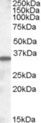
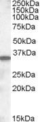
- Western Blot staining of Mouse Small Intestine lysate using PA5-18990 at a concentration of 0.5 ug/ml, the primary antibody incubation was 1 hour and the detection method was chemiluminescence.
Supportive validation
- Submitted by
- Invitrogen Antibodies (provider)
- Main image
- Experimental details
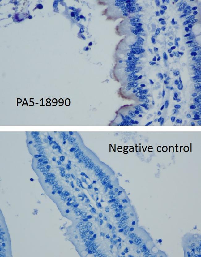
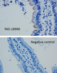
- Immunohistochemistry analysis of SLC10A2 Polyclonal Antibody (Product # PA5-18990) was performed on a pig ileum fixed in 10% formalin for 24 hours. To expose target proteins, antigen retrieval was performed by heating tissues in a pressure cooker for 10 minutes in 10mM sodium citrate buffer (pH 6.0). Following antigen retrieval, endogenous peroxidases were blocked with 3% hydrogen peroxide for 10 min at room temperature. Tissue slides were washed with deionized water and TBST, and then blocked in a protein free blocking buffer diluted 1:10 for 7 min at room temperature. Tissues were incubated with a polyclonal SLC10A2 antibody (Product # PA5-18990) diluted 1:100 in a blocking serum or goat IgG as a negative control for 1 hour at room temperature in a humidified chamber. Tissues were washed extensively in TBST and then followed by colorimetric detection using a DAB kit. Tissues were counterstained with hematoxylin and dehydrated with ethanol and xylene to prep for mounting. Images were taken at 40X magnification. In the image, immunoreactivity is observed in the brush border of enterocytes in porcine ileum using the described technique for immunohistochemistry. Data courtesy of Antibody Data Exchange Program.