Explore
 Validate
 Learn
Learn Western blot
Antibody data
- Antibody Data
- Antigen structure
- References [0]
- Comments [0]
- Validations
- Western blot [3]
- Immunocytochemistry [1]
- Immunohistochemistry [1]
Submit
Validation data
Reference
Comment
Report error
- Product number
- PA5-19148 - Provider product page

- Provider
- Invitrogen Antibodies
- Product name
- SATB1 Polyclonal Antibody
- Antibody type
- Polyclonal
- Antigen
- Synthetic peptide
- Description
- This antibody is predicted to react with bovine, mouse and rat based on sequence homology.
- Reactivity
- Human
- Host
- Goat
- Isotype
- IgG
- Vial size
- 100 µg
- Concentration
- 0.5 mg/mL
- Storage
- -20° C, Avoid Freeze/Thaw Cycles
No comments: Submit comment
Supportive validation
- Submitted by
- Invitrogen Antibodies (provider)
- Main image
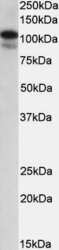
- Experimental details
- Western Blot staining of MOLT4 cell lysate using Product # PA5-19148 at a concentration of 0.3 µg/mL, the primary antibody incubation was 1 hour and the detection method was chemiluminescence.
- Submitted by
- Invitrogen Antibodies (provider)
- Main image
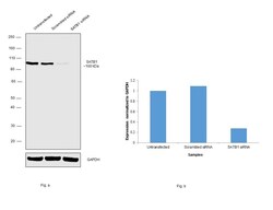
- Experimental details
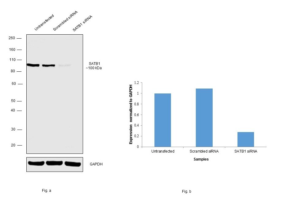
- Knockdown of SATB1 was achieved by transfecting IMR-32 with SATB1 specific siRNAs (Silencer® select Product # s12481, s12479). Western blot analysis (Fig. a) was performed using whole cell extracts from the SATB1 knockdown cells (Lane 3), non-specific scrambled siRNA transfected cells (Lane 2) and untransfected cells (Lane 1). The blot was probed with SATB1 Polyclonal Antibody (Product # PA5-19148, 0.3ug/ml) and Rabbit anti-Goat IgG (H+L) Superclonal™ Recombinant Secondary Antibody, HRP (Product # A27014, 1:4000 dilution). Densitometric analysis of this western blot is shown in histogram (Fig. b). Decrease in signal upon siRNA mediated knock down confirms that antibody is specific to SATB1.
- Submitted by
- Invitrogen Antibodies (provider)
- Main image
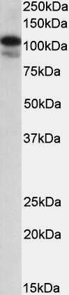
- Experimental details
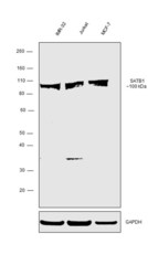
- Western blot was performed using Anti-SATB1 Polyclonal Antibody (Product # PA5-19148) and a ~100 kDa band corresponding to SATB1 was observed in IMR-32, Jurkat and MCF-7 cell lines. Whole cell extracts (30 µg lysate) of IMR-32 (Lane 1), Jurkat (Lane 2) and MCF-7 (Lane 3) were electrophoresed using NuPAGE™ 10% Bis-Tris Protein Gel (Product # NP0302BOX). Resolved proteins were then transferred onto a nitrocellulose membrane (Product # IB23001) by iBlot® 2 Dry Blotting System (Product # IB21001). The blot was probed with the primary antibody (0.3 µg/mL) and detected by chemiluminescence with Rabbit anti-Goat IgG (H+L), Superclonal™ Recombinant Secondary Antibody, HRP (Product # A27014, 1:4000 dilution) using the iBright FL 1000 (Product # A32752). Chemiluminescent detection was performed using Novex® ECL Chemiluminescent Substrate Reagent Kit (Product # WP20005).

Supportive validation
- Submitted by
- Invitrogen Antibodies (provider)
- Main image

- Experimental details
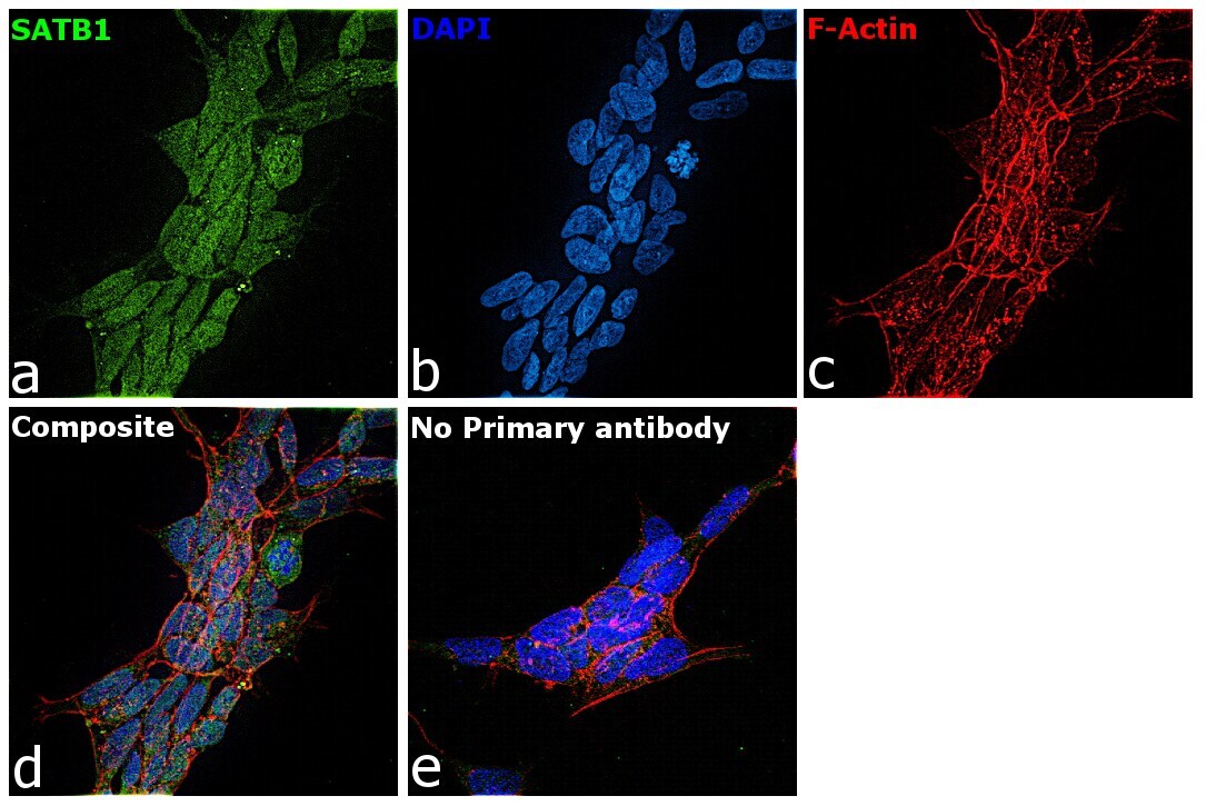
- Immunofluorescence analysis of HUR was performed using 70% confluent log phase IMR-32 cells. The cells were fixed with 4% paraformaldehyde for 10 minutes, permeabilized with 0.1% Triton™ X-100 for 15 minutes, and blocked with 2% BSA for 1 hour at room temperature. The cells were labeled with HUR Rabbit Polyclonal Antibody (Product # PA5-19148) at 5 µg/mL in 0.1% BSA, incubated at 4 degree celsius overnight and then labeled with Rabbit anti-Goat IgG (H+L) Cross-Adsorbed Secondary Antibody, Alexa Fluor 488 (Product # A-11078) at a dilution of 1:2000 for 45 minutes at room temperature (Panel a: green). Nuclei (Panel b: blue) were stained with ProLong™ Diamond Antifade Mountant with DAPI (Product # P36962). F-actin (Panel c: red) was stained with Rhodamine Phalloidin (Product # R415). Panel d represents the merged image showing Nuclear localization. Panel e represents control cells with no primary antibody to assess background. The images were captured at 60X magnification.
Supportive validation
- Submitted by
- Invitrogen Antibodies (provider)
- Main image
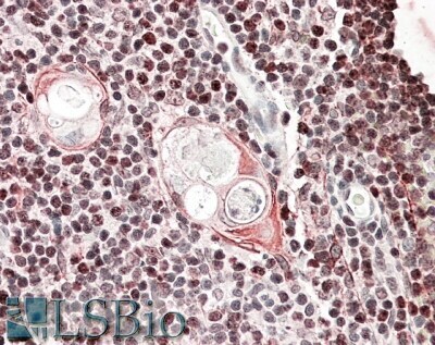
- Experimental details
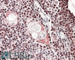
- Immunohistochemical analysis of SATB1 in paraffin embedded human thymus using a SATB1 polyclonal antibody (Product #PA5-19148) at a concentration of 3.8 µg/mL. Steamed antigen retrieval was performed with pH 6 buffered citrate. Samples were then stained with alkaline phosphatase.